Images & credits
NeuroScience Decoded uses 86 photographs and historical images from Wikimedia Commons — each downloaded, downscaled and re-hosted here — plus 13 original teaching diagrams and 98 pieces of original cover art, released under CC0. Full attribution for every image is below.
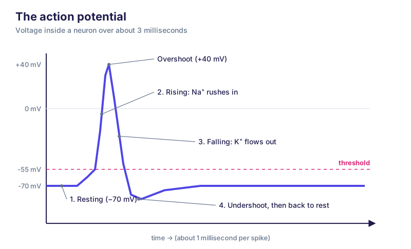
Original teaching diagram: action potential.
NeuroScience Decoded original diagram | NeuroScience Decoded | CC0 1.0
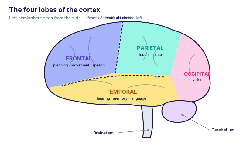
Original teaching diagram: brain lobes.
NeuroScience Decoded original diagram | NeuroScience Decoded | CC0 1.0
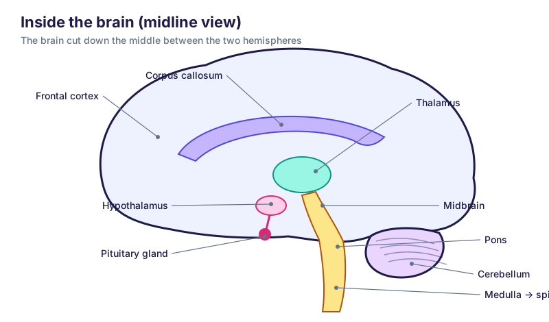
Original teaching diagram: brain sagittal.
NeuroScience Decoded original diagram | NeuroScience Decoded | CC0 1.0

Original teaching diagram: brain waves.
NeuroScience Decoded original diagram | NeuroScience Decoded | CC0 1.0

Original teaching diagram: memory types.
NeuroScience Decoded original diagram | NeuroScience Decoded | CC0 1.0
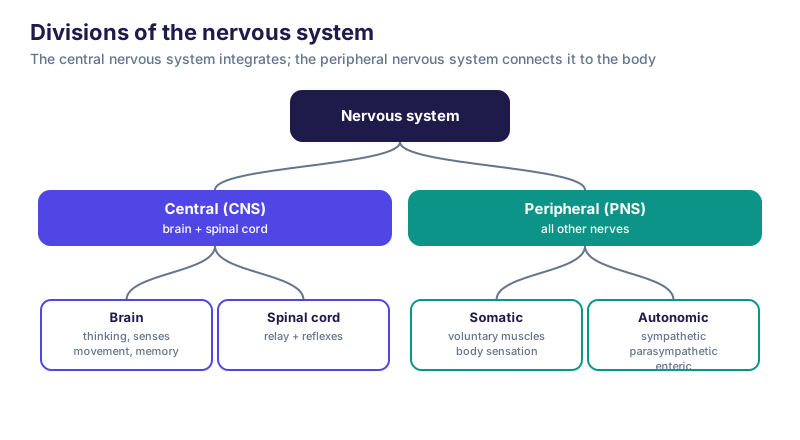
Original teaching diagram: nervous system divisions.
NeuroScience Decoded original diagram | NeuroScience Decoded | CC0 1.0
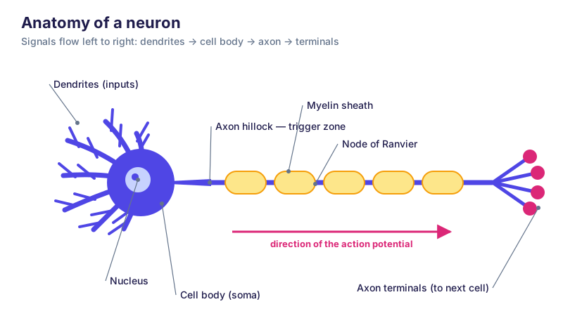
Original teaching diagram: neuron anatomy.
NeuroScience Decoded original diagram | NeuroScience Decoded | CC0 1.0

Original teaching diagram: neurotransmitters.
NeuroScience Decoded original diagram | NeuroScience Decoded | CC0 1.0
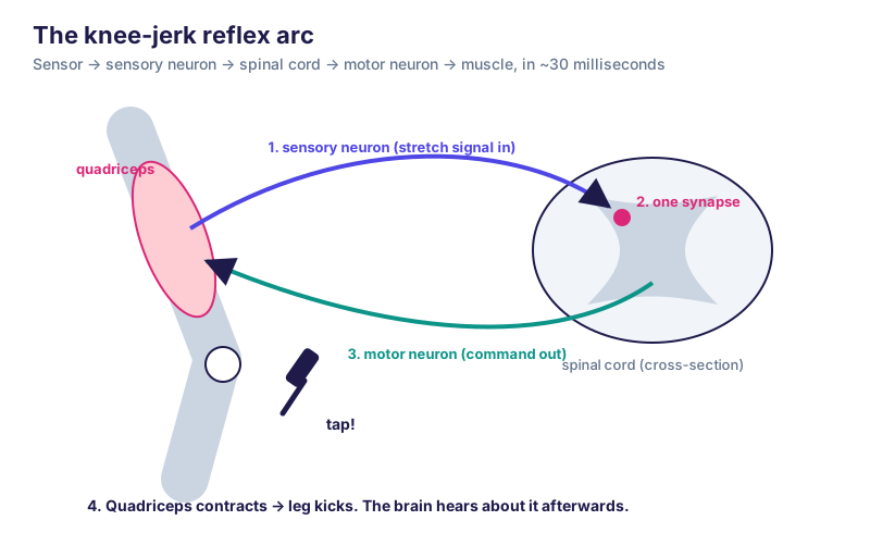
Original teaching diagram: reflex arc.
NeuroScience Decoded original diagram | NeuroScience Decoded | CC0 1.0
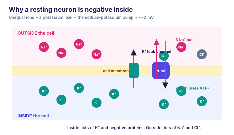
Original teaching diagram: resting potential.
NeuroScience Decoded original diagram | NeuroScience Decoded | CC0 1.0
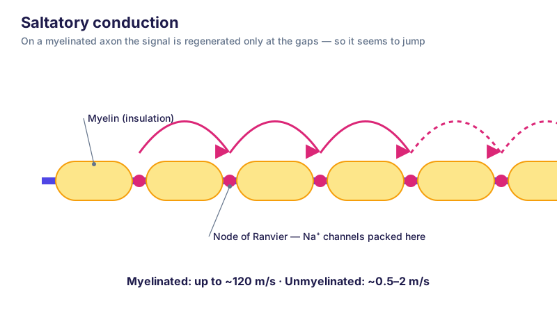
Original teaching diagram: saltatory conduction.
NeuroScience Decoded original diagram | NeuroScience Decoded | CC0 1.0
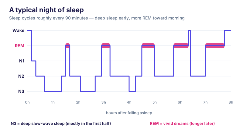
Original teaching diagram: sleep cycle.
NeuroScience Decoded original diagram | NeuroScience Decoded | CC0 1.0
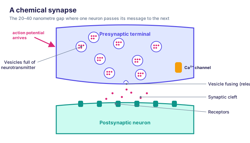
Original teaching diagram: synapse.
NeuroScience Decoded original diagram | NeuroScience Decoded | CC0 1.0
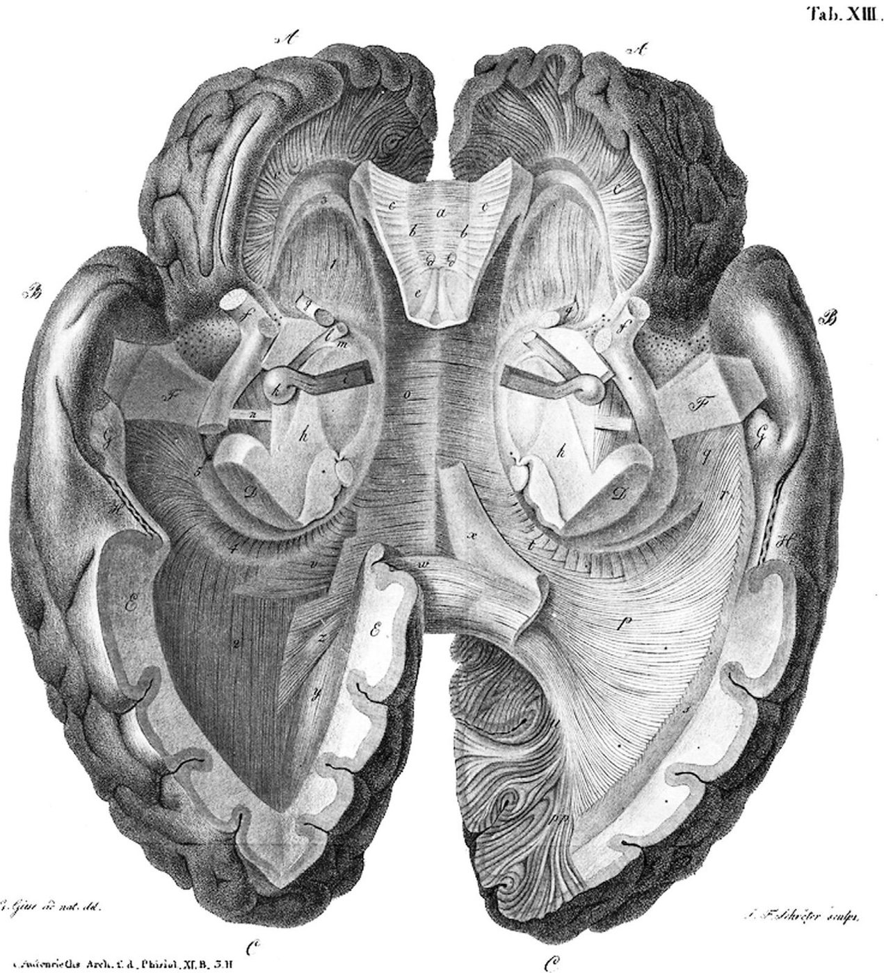
Johann Christian Reil’s depiction of his gross dissection of the human brain viewed from below. (Reil, 1812, volume XI, Tafel XIII)
Wikimedia Commons — File:Dissection of the human brain, Johann Christian Reil, 1812.jpg | Johann Christian Reil | Public domain
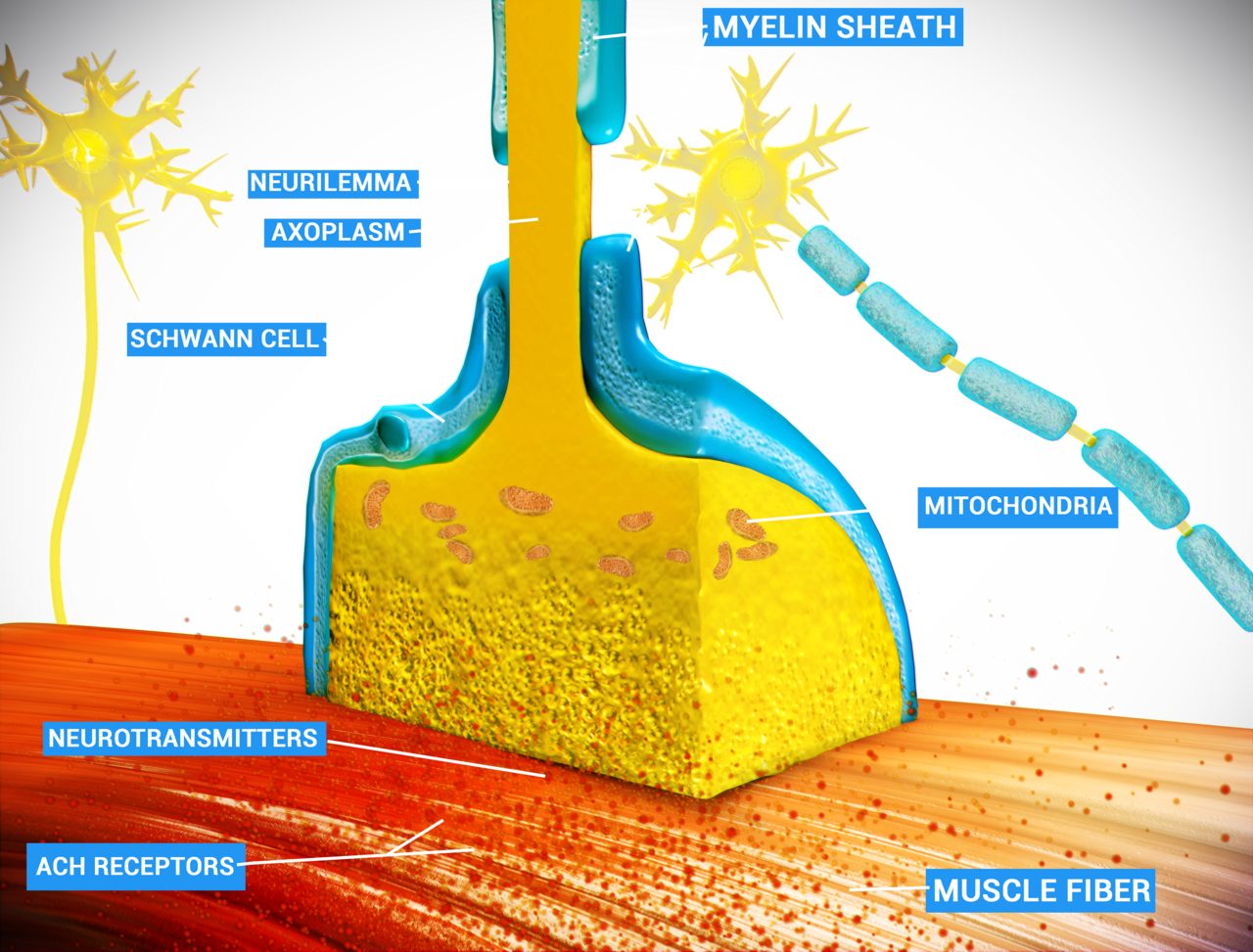
A neuromuscular junction (or myoneural junction) is a chemical synapse formed by the contact between a motor neuron and a muscle fiber. It is at the neuromuscular junction that a motor neuron is able to transmit a signal
Wikimedia Commons — File:Neuro Muscular Junction.png | Doctor Jana | CC BY 4.0
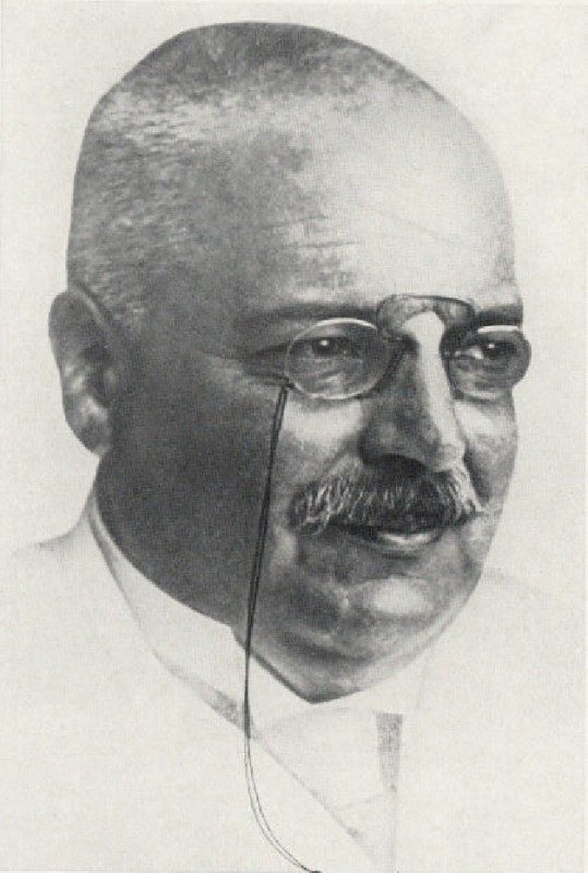
Alois Alzheimer
Wikimedia Commons — File:Alois Alzheimer 003.jpg | uncredited | Public domain
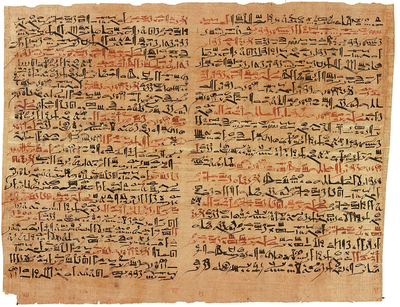
The Edwin Smith papyrus, the world's oldest surviving surgical document. Written in hieratic script in ancient Egypt around 1600 B.C., the text describes anatomical observations and the examination, diagnosis, treatment,
Wikimedia Commons — File:Edwin Smith Papyrus v2.jpg | Jeff Dahl | Public domain
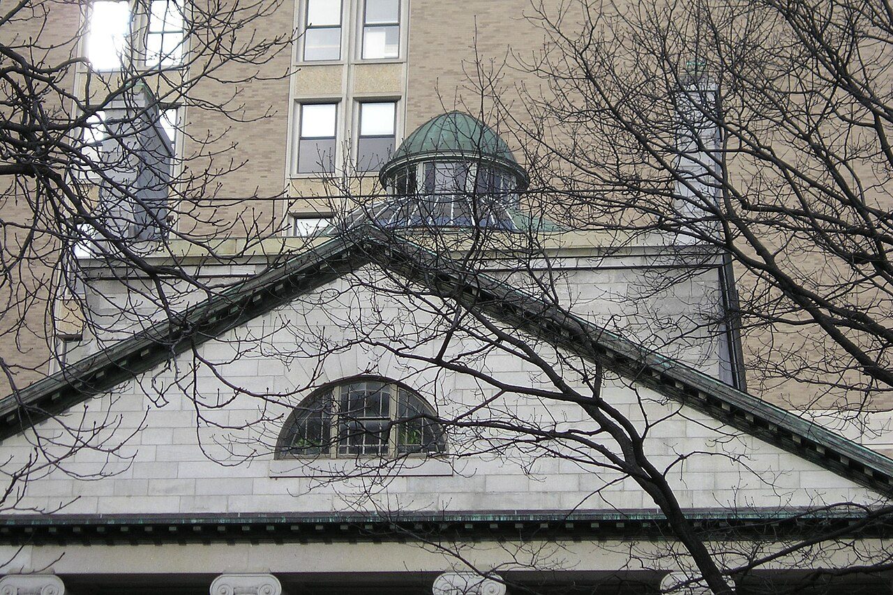
The Ether Dome atop the Bulfinch Building at Massachusetts General Hospital in downtown Boston as photographed from the grounds on 29 January 2007
Wikimedia Commons — File:MGH Ether Dome Outside 29Jan2008.jpg | Adam Lenhardt | CC BY-SA 3.0
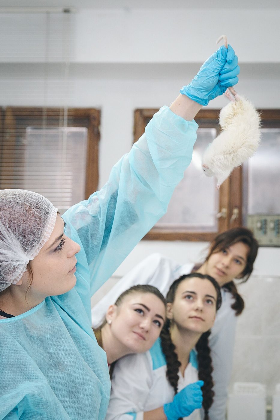
Students of the V.I. Vernadsky Crimean Federal University carry out experiments on laboratory mice
Wikimedia Commons — File:Researcher holding a lab mouse - Crimean Federal University - ANDY7392.jpg | Andrey Chernogorodov | CC BY 4.0
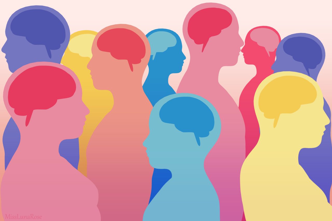
Everyone's brain works a little differently. According to the neurodiversity paradigm, this isn't necessarily a bad thing. Society can benefit from the strengths and accommodate the weaknesses of each person
Wikimedia Commons — File:Neurodiversity Crowd 2.png | MissLunaRose12 | CC BY-SA 4.0
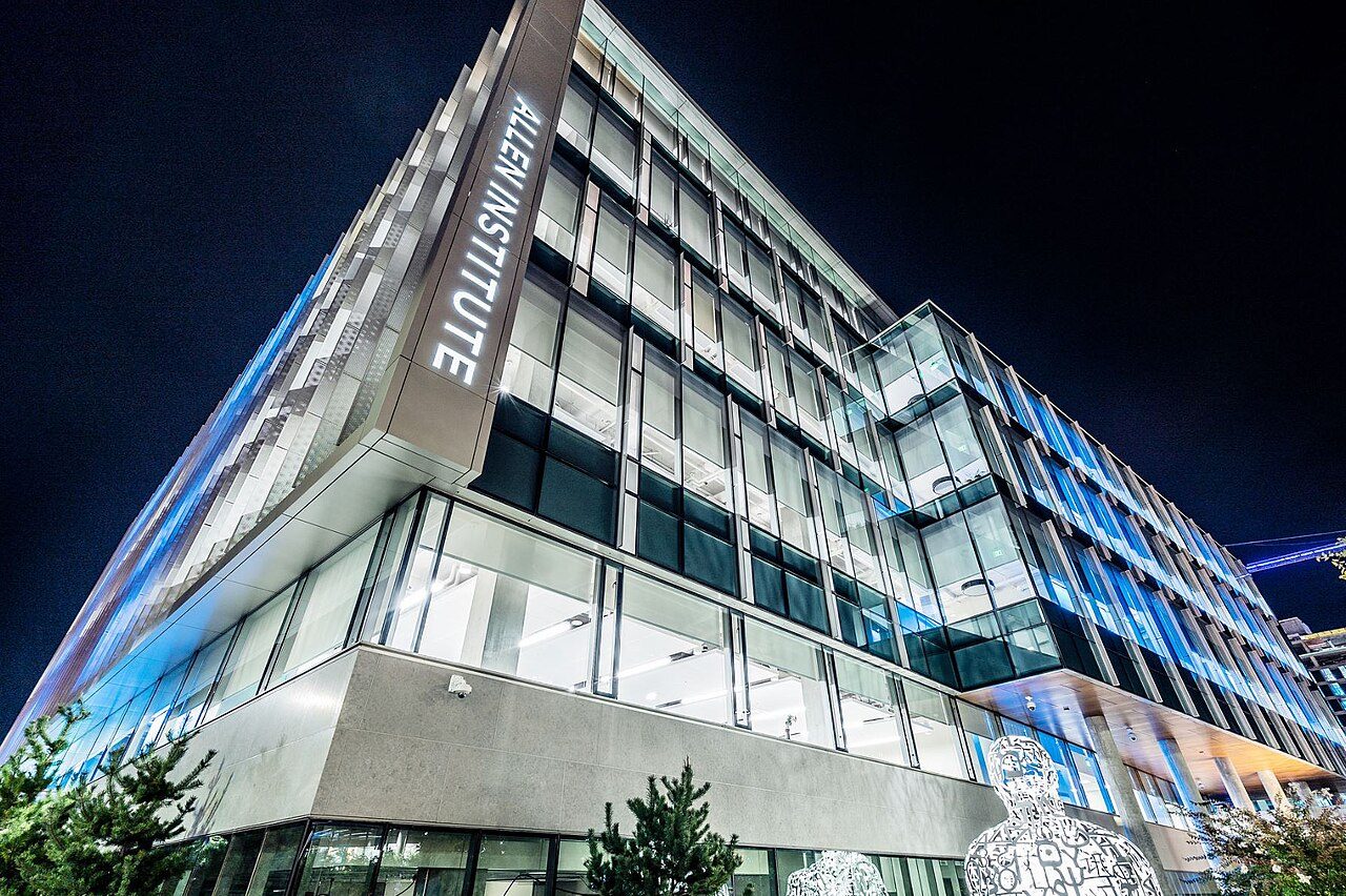
Allen Institute Headquarters
Wikimedia Commons — File:Allen Institute Headquarters at night.jpg | Nick.hawley | CC BY-SA 4.0
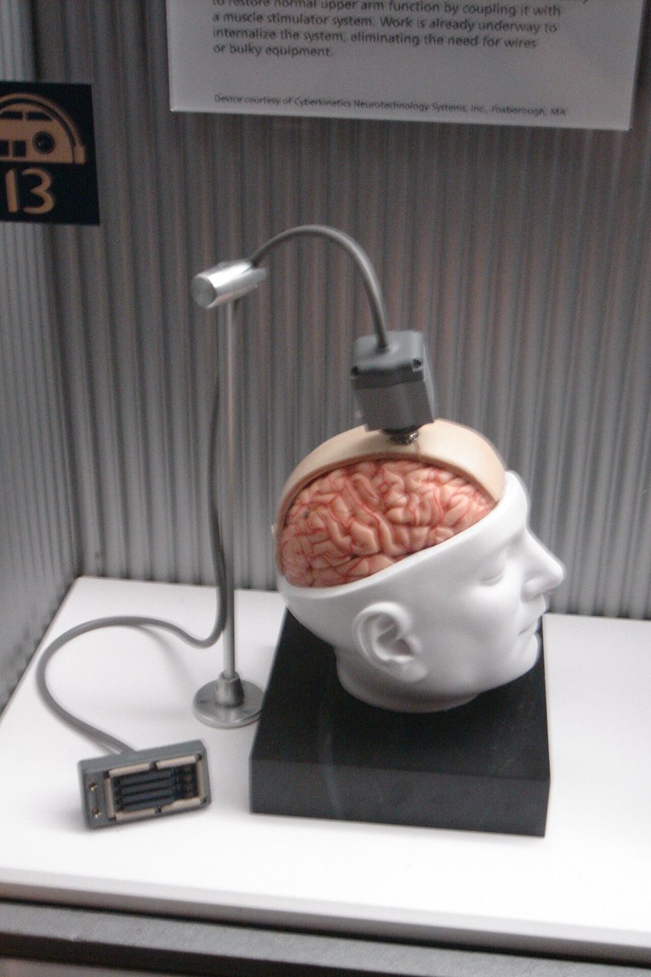
This is a photo of a dummy BrainGate interface which was at the Star Wars exhibition at the Boston Science Museum in October 2005. I took it myself
Wikimedia Commons — File:BrainGate.jpg | PaulWicks at English Wikipedia | Public domain
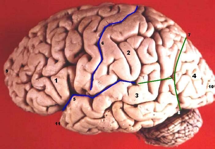
Human brain lateral view - Lobes Lobus frontalis Lobus parietalis Lobus temporalis Lobus occipitalis Sulcus lateralis Sulcus centralis Sulcus parietooccipitalis Incisura preoccipitalis Polus frontalis Polus occipitalis P
Wikimedia Commons — File:Human brain lateral view description.JPG | John A Beal, PhD Dep't. of Cellular Biology & Anatomy, Louisiana State University Health Sciences Center Shreveport | CC BY 2.5
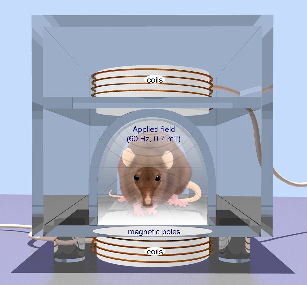
Repetitive transcranial magnetic stimulation (rTMS) is a technique for noninvasive stimulation of the adult brain. Stimulation is produced by generating a brief, high-intensity magnetic field by passing a brief electric
Wikimedia Commons — File:Repetitive transcranial magnetic stimulation (rTMS) is a technique for noninvasive stimulation of the adult brain.jpg | Oscar Arias-Carrión | CC BY 2.0
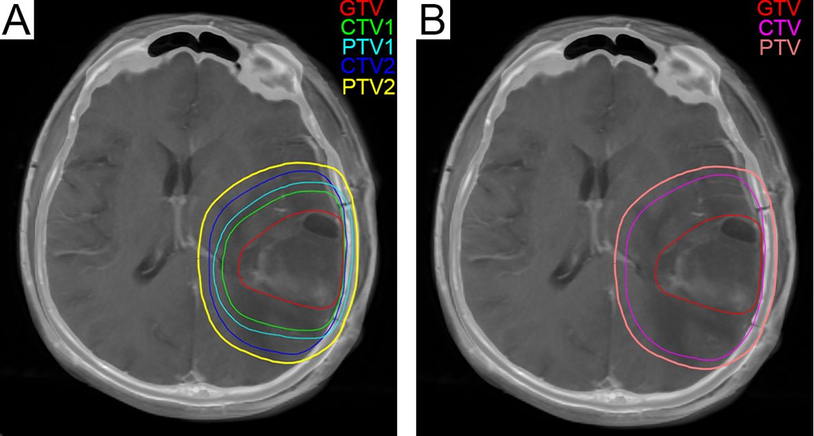
(A) The green line represents CTV1 in the radiation treatment planning in our center, and 204.6 cm3 was exposed to a dose of 60 Gy. (B) The fuchsia contour represents the CTV in the EORTC radiotherapy plan, and 347.1 cm3
Wikimedia Commons — File:Glioblastoma radiation treatment plan.png | Lin Zheng, Zhi-Rui Zhou, QianQian Yu, Minghan Shi, Yang Yang, Xiaofeng Zhou, Chao Li, Qichun Wei | CC BY 4.0
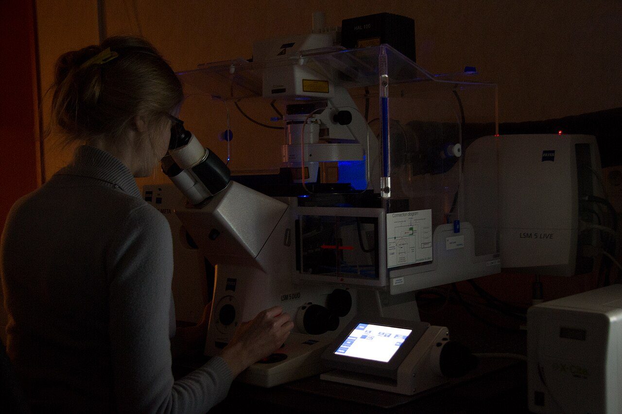
Konfokaalmikroskoobi kasutamine neuronite uurimiseks Biomeedikumis, Tartu Ülikool
Wikimedia Commons — File:Neuroscience 2.jpg | Priit Tiganik | CC BY-SA 3.0
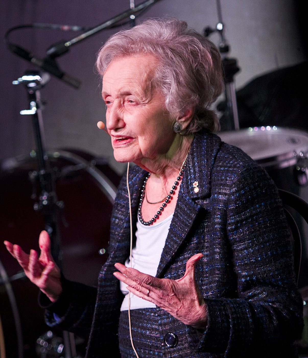
Neuropsychologist Brenda Milner at TEDxMcGill, 2011. Photo by Eva Blue
Wikimedia Commons — File:Brenda Milner.jpg | Eva Blue | CC BY 2.0
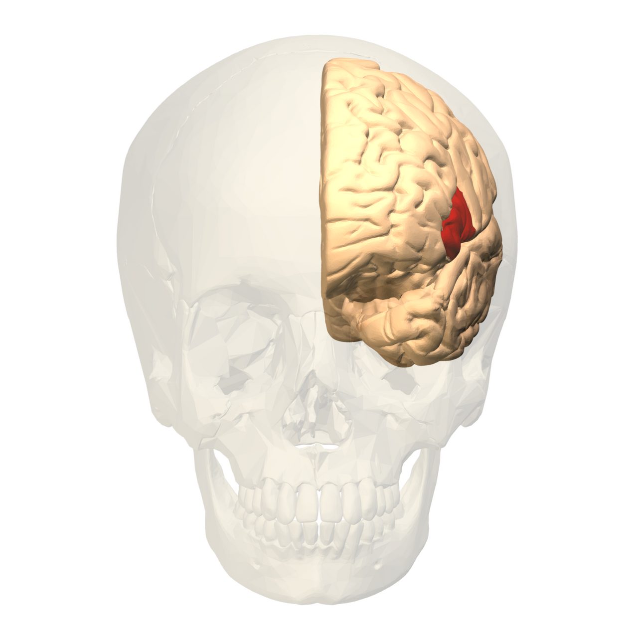
Broca's area (shown in red).Colored region is pars opercularis and pars triangularis of the inferior frontal gyrus. Broca's area is now typically defined in terms of the pars opercularis and pars triangularis of the infe
Wikimedia Commons — File:Broca's area - anterior view.png | Polygon data were generated by Database Center for Life Science(DBCLS)[2] | CC BY-SA 2.1 jp
A photo of a cup of coffee
Wikimedia Commons — File:A small cup of coffee.JPG | Julius Schorzman | CC BY-SA 2.0
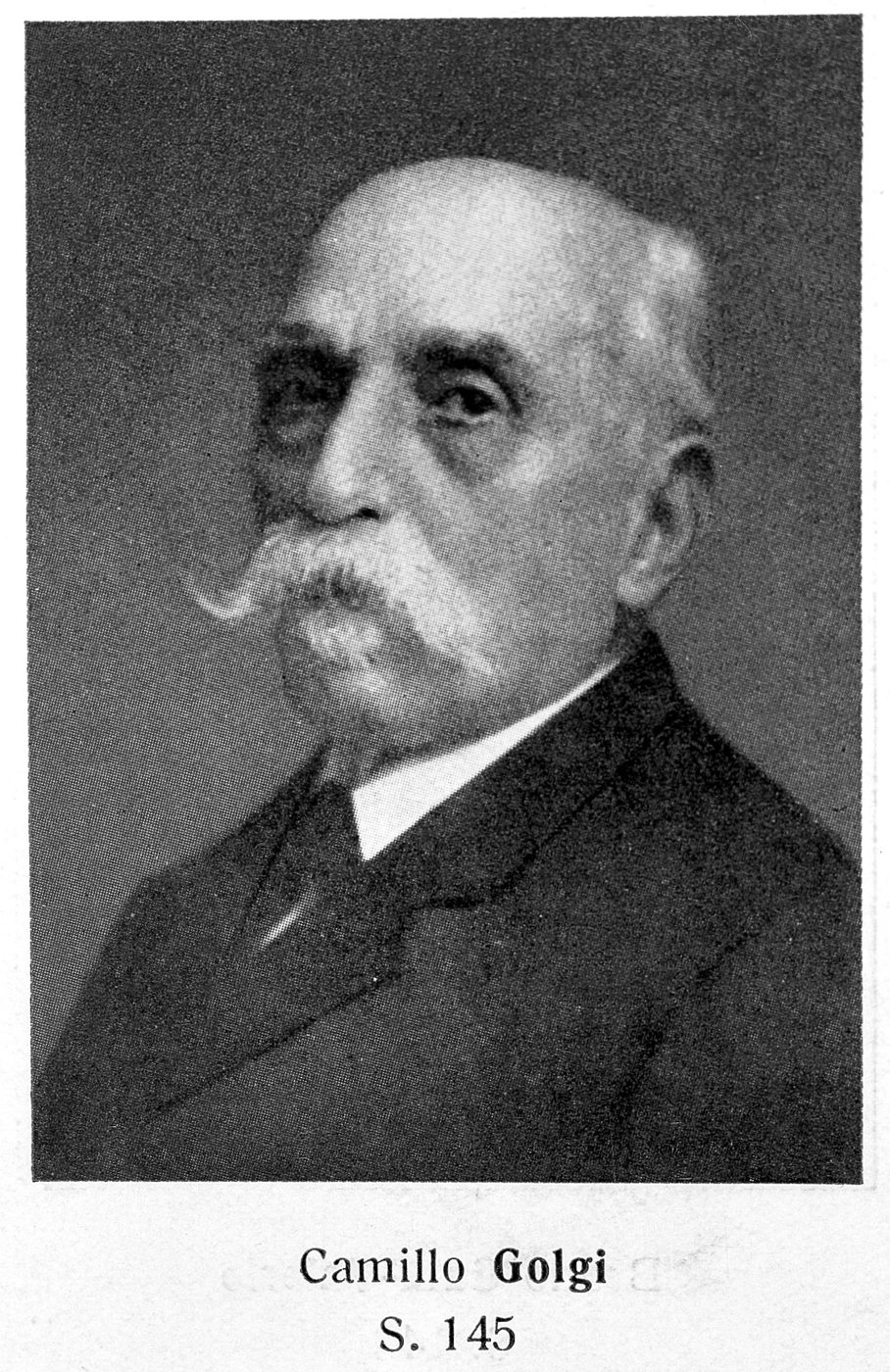
Portrait of Camillo Golgi. General Collections Keywords: Camillo Golgi
Wikimedia Commons — File:Portrait of Camillo Golgi. Wellcome L0011979.jpg | Unknown | CC BY 4.0
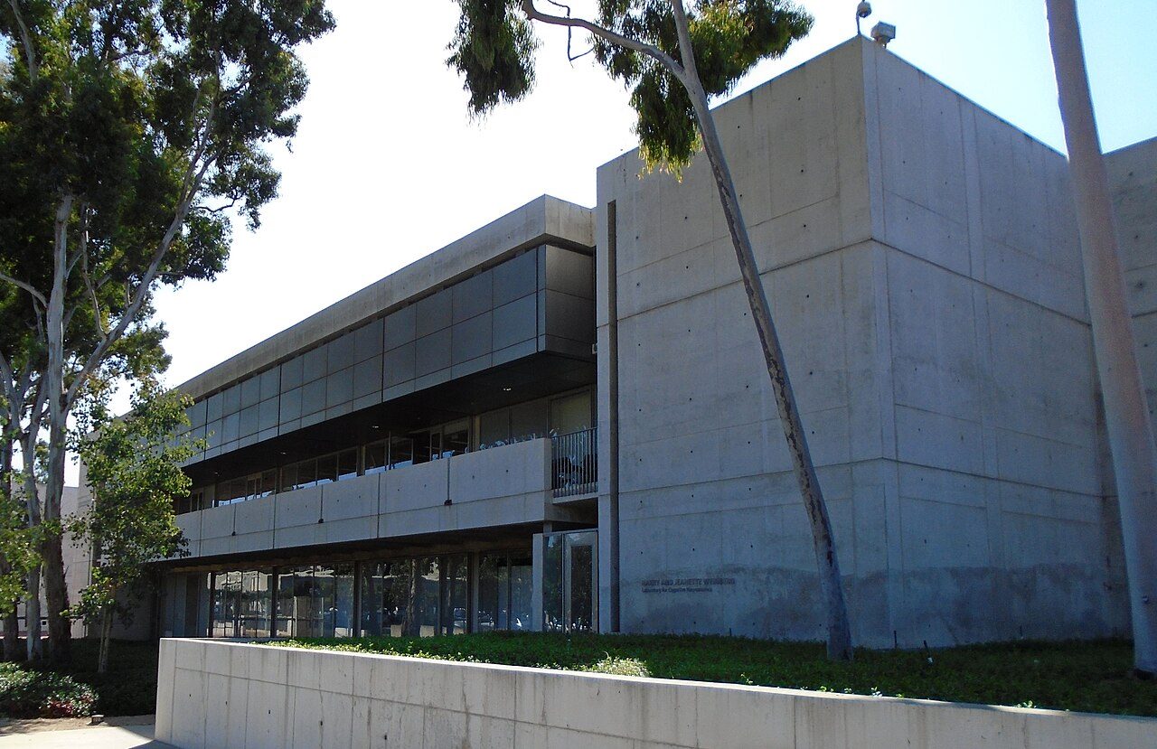
The Henry and Jeanette Weinberg Laboratory for Cognitive Neuroscience in the north wing of the Salk Institute at 10010 North Torrey Pines Road in La Jolla, San Diego, California. The institute was founded in 1960 by Jona
Wikimedia Commons — File:2019 Salk Institute Henry and Jeanette Weinberg Laboratory for Cognitive Neuroscience (north wing).jpg | Beyond My Ken | CC BY-SA 4.0
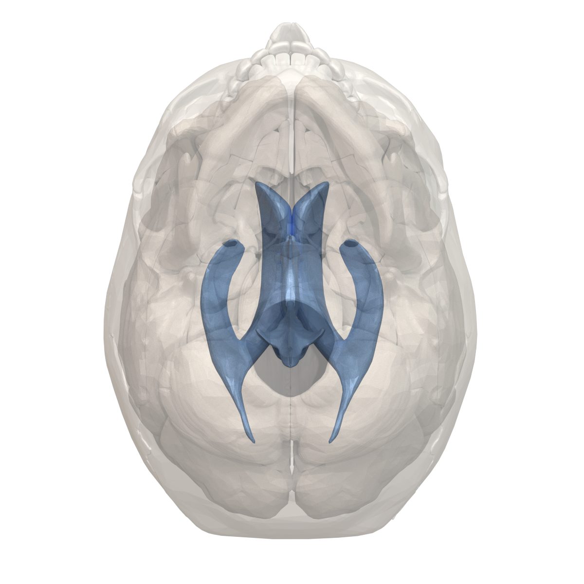
Human ventricular system (shown in blue) seen from the bottom
Wikimedia Commons — File:Human ventricular system - bottom view.png | BodyParts3D[1] by DBCLS | CC BY-SA 2.1 jp
Sunrise at Viru Bog, Estonia
Wikimedia Commons — File:Sunrise at viru bog.jpg | Abrget47j | CC BY-SA 3.0
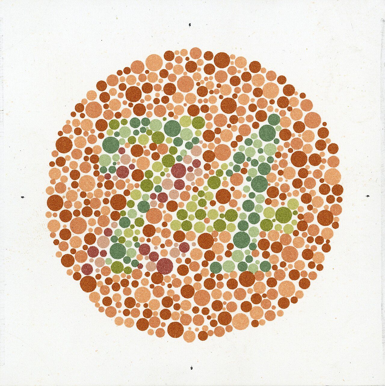
Eight Ishihara charts for testing colour blindness, Europe, 1917-1959 Colour blindness is tested using these eight placards. They are known as Ishihara charts. They are named after their inventor, Japanese ophthalmologis
Wikimedia Commons — File:Eight Ishihara charts for testing colour blindness, Europe Wellcome L0059158.jpg | Unknown | CC BY 4.0
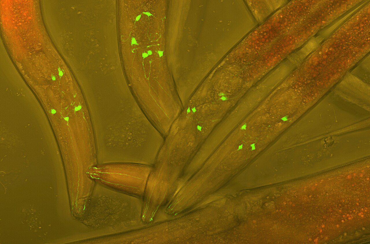
Alive Caenorhabditis elegans worms, who have GFP (green fluorescent protein) inserted into their neurons to visualize neural development in a living worm. Differential interference contrast micrograph + fluorescence
Wikimedia Commons — File:Varbuss.jpg | Heiti Paves | CC BY-SA 3.0
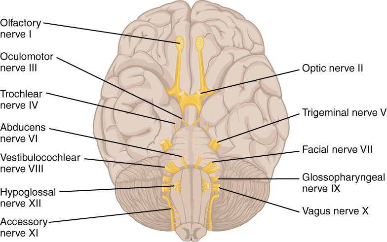
Version 8.25 from the Textbook OpenStax Anatomy and Physiology Published May 18, 2016
Wikimedia Commons — File:1320 The Cranial Nerves.jpg | OpenStax | CC BY 4.0
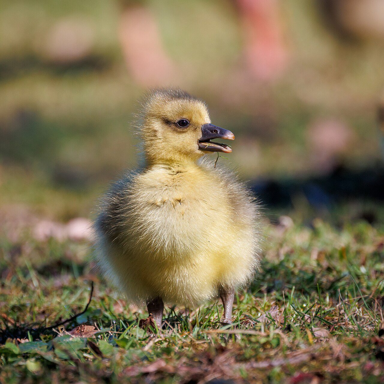
Greylag gosling
Wikimedia Commons — File:Greylag goose gosling.jpg | Stig Nygaard | CC BY 2.0
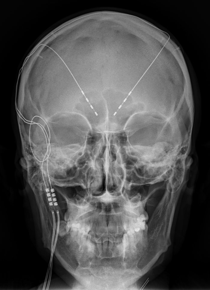
Frontal X-ray of the head: Deep brain stimulation in obsessive–compulsive disorder (Medtronic 3391). Men 42 years old, surgery in 2013
Wikimedia Commons — File:X-ray of deep brain stimulation in OCD, F.png | Jmarchn | CC BY-SA 3.0
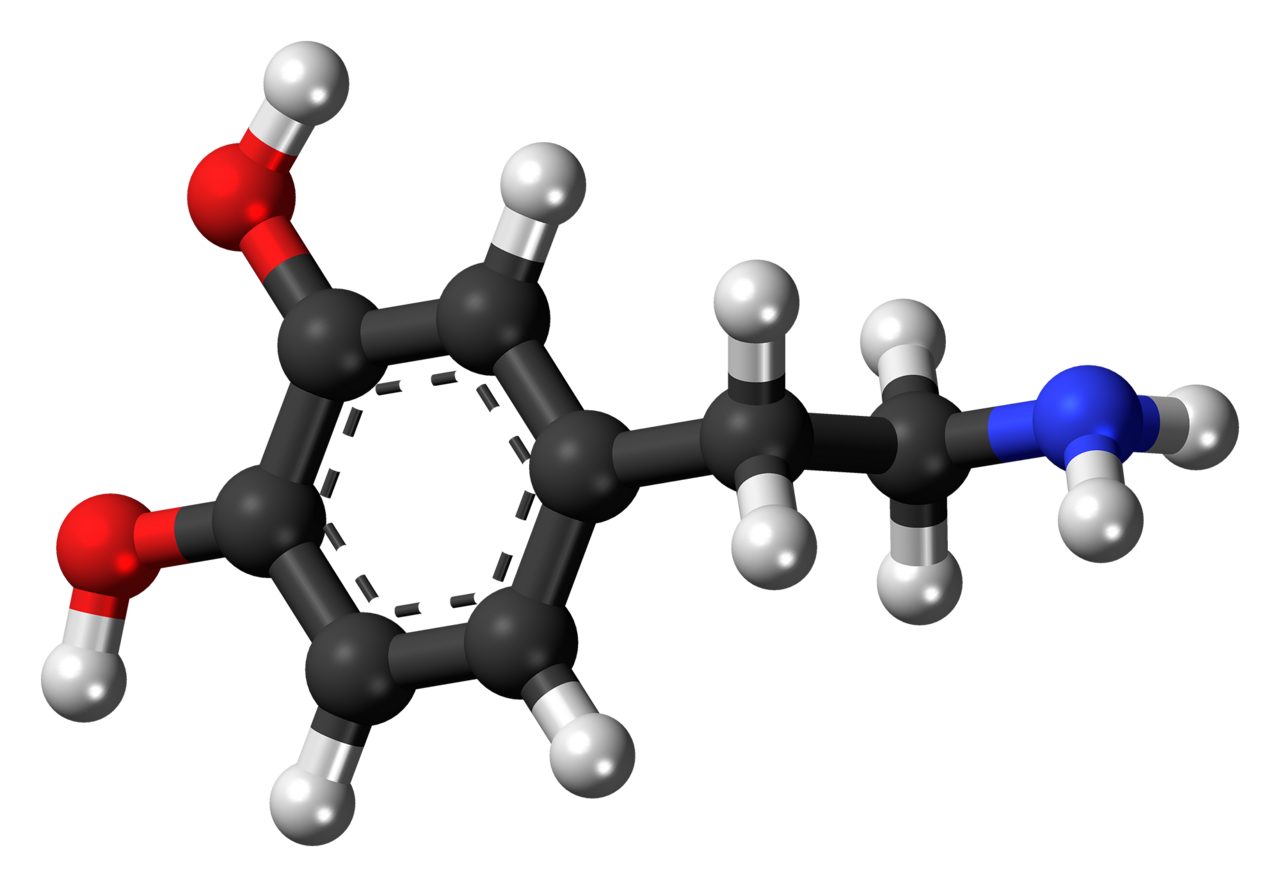
Ball-and-stick model of the dopamine molecule, a neurotransmitter that affects the brain's reward and pleasure centers. Colour code: Carbon, C: black Hydrogen, H: white Oxygen, O: red Nitrogen, N: blue
Wikimedia Commons — File:Dopamine 3D ball.png | Jynto (talk) | CC0
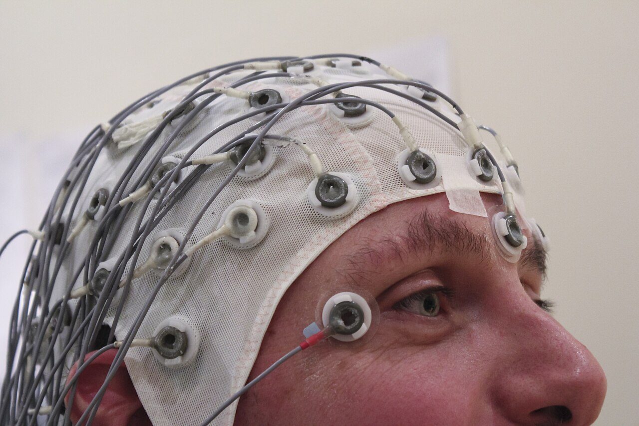
A cap holds electrodes in place while recording an EEG
Wikimedia Commons — File:EEG Recording Cap.jpg | Chris Hope | CC BY 2.0
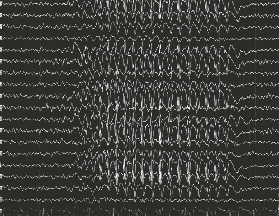
The EEG signature of absence epilepsy is the generalized 3 Hz spike-wave discharge
Wikimedia Commons — File:EEG Absence seizure.png | Bromfield EB, Cavazos JE, Sirven JI, editors | CC BY-SA 4.0

A Marine of the United States Marine Corps runs through a creek. Original caption: "Lance Cpl. Anthony M. Madonia emerges from the water during the swimming portion of the triathlon. Marines and Sailors of Marine Securit
Wikimedia Commons — File:Soldier running in water.jpg | Cpl. Earnest J. Barnes | Public domain
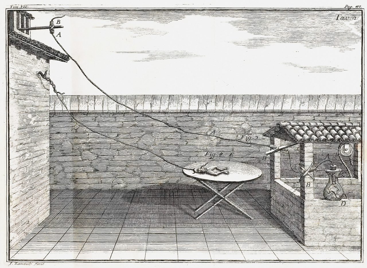
Diagram of Galvani's experiment in frog legs
Wikimedia Commons — File:Galvani frog legs experiment setup.png | Luigi Galvani | Public domain
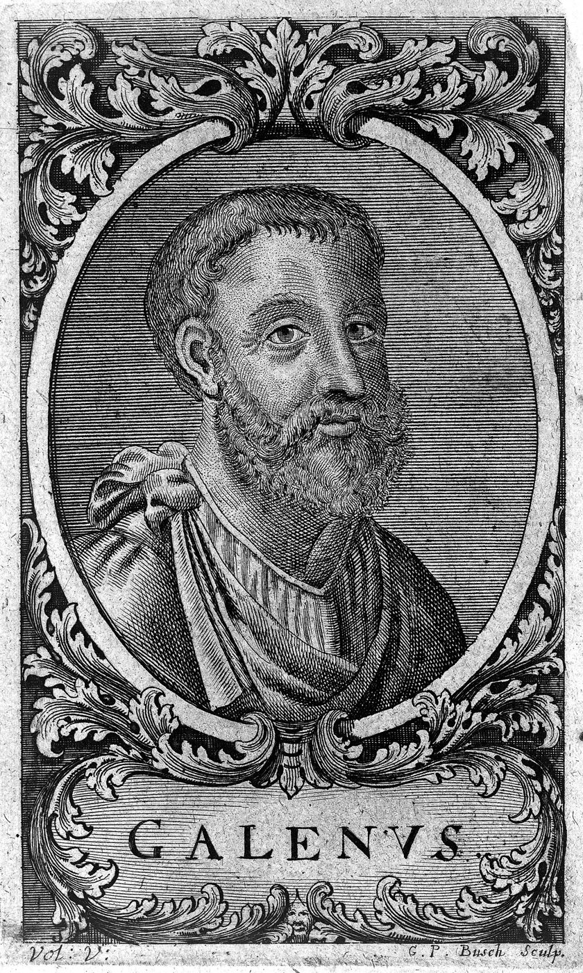
GALEN {c. 130-200 A.D.} Engraving: 'portrait' of Galen, head and shoulders; by G. P. Busch, n.d. S 10 S 10 Iconographic Collections Keywords: burgess, portraits, 1069.3; galen {c. 130-200 a.d.}; Portraits
Wikimedia Commons — File:Engraving; 'portrait' of Galen, head and Wellcome L0005549.jpg | Unknown | CC BY 4.0
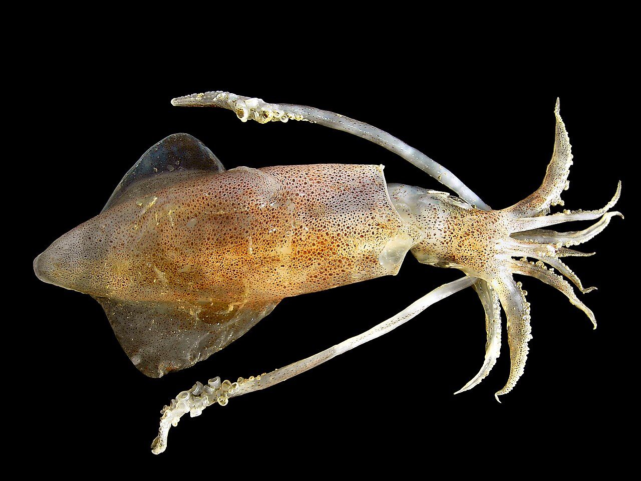
Common squid from the Belgian continental shelf. Picture taken in the lab on board of the RV Belgica, of a live specimen to preserve colour and structure of chromophores. Mantle: length= 138 mm, width= 39 mm
Wikimedia Commons — File:Loligo vulgaris.jpg | Hans Hillewaert | CC BY-SA 4.0
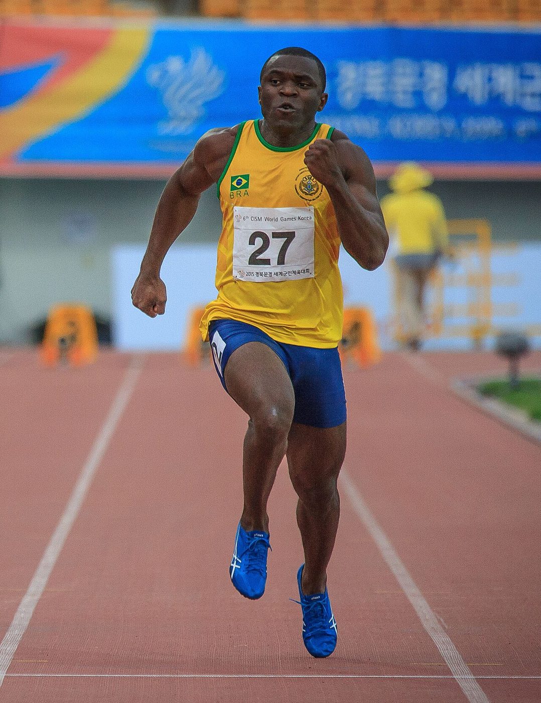
José Carlos Moreira at the 2015 Military World Games
Wikimedia Commons — File:José Carlos Moreira 2015.jpg | Johnson Barros | CC BY 2.0
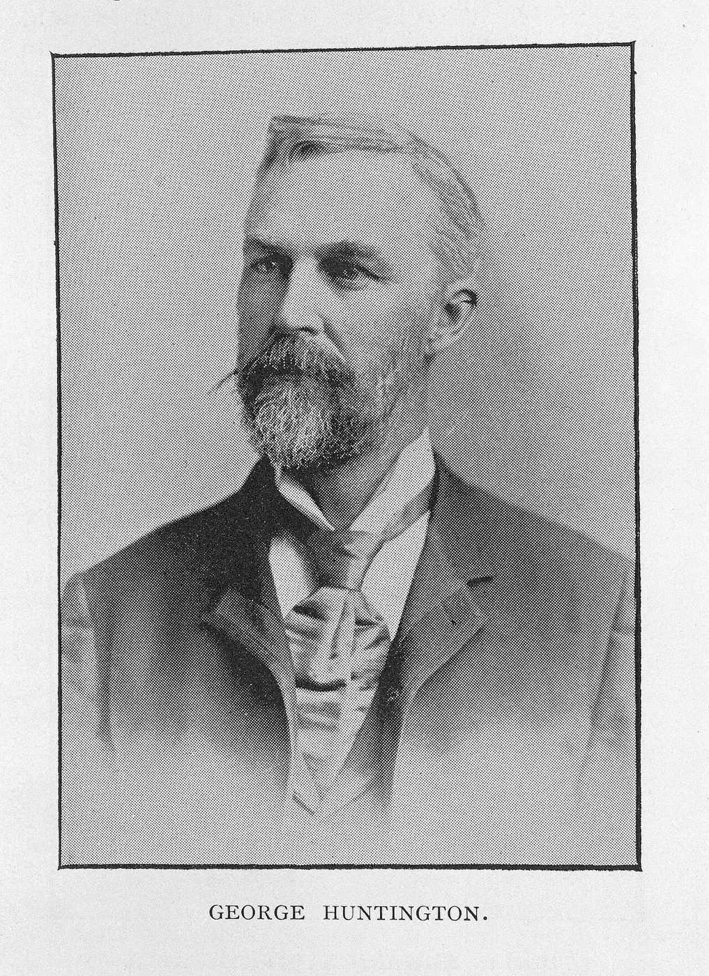
Portrait of George Huntington General Collections Keywords: gc; surgeons; Physicians
Wikimedia Commons — File:Portrait of George Huntington Wellcome L0029545.jpg | Unknown | CC BY 4.0
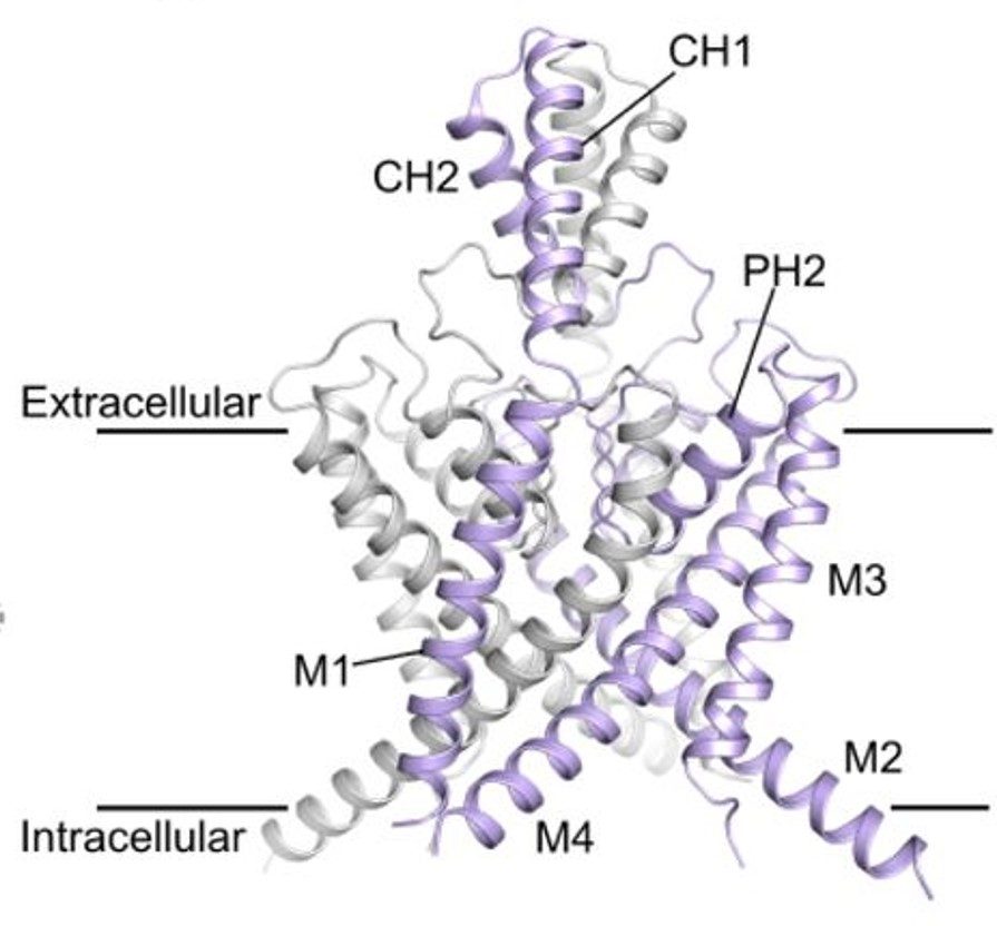
Ribbon structure of homodimeric two-pore potassium channel K2P13 (THIK-1). Subunits are colored in gray and purple. Transmembrane helices M1-M4, pore helix PH2 (PH1 not shown), and cap helices CH1-CH2 are labeled. Horizo
Wikimedia Commons — File:Two-pore domain potassium channel K2P13.jpg | Karin EJ Rödström, Bisher Eymsh, Peter Proks, Mehtab S Hayre, Christian Madry, Anna Rowland, Simon Newstead, Thomas Bauk | CC BY 4.0

A Ghanaian sign language interpreter at a Wikipedia training at the Special Education Department of the University of Education, Winneba
Wikimedia Commons — File:Ghanaian sign language interpreter working at University of Education Winneba.jpg | Uprising Man | CC BY-SA 4.0
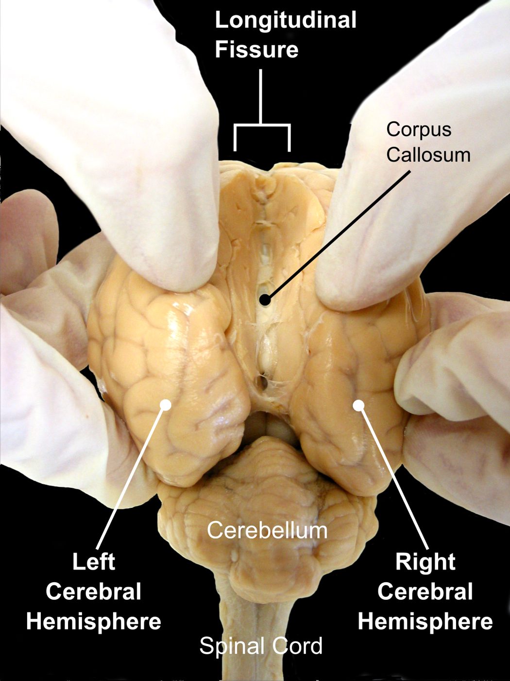
Sheep Brain. Opening longitudinal fissure, the fissure devide left and right cerebral hemispheres
Wikimedia Commons — File:Sheep Brain Dissection 2 - black background with labels.png | Original photo is by aaronbflickr. Making background black and adding labels by was_a_bee | CC BY 2.0
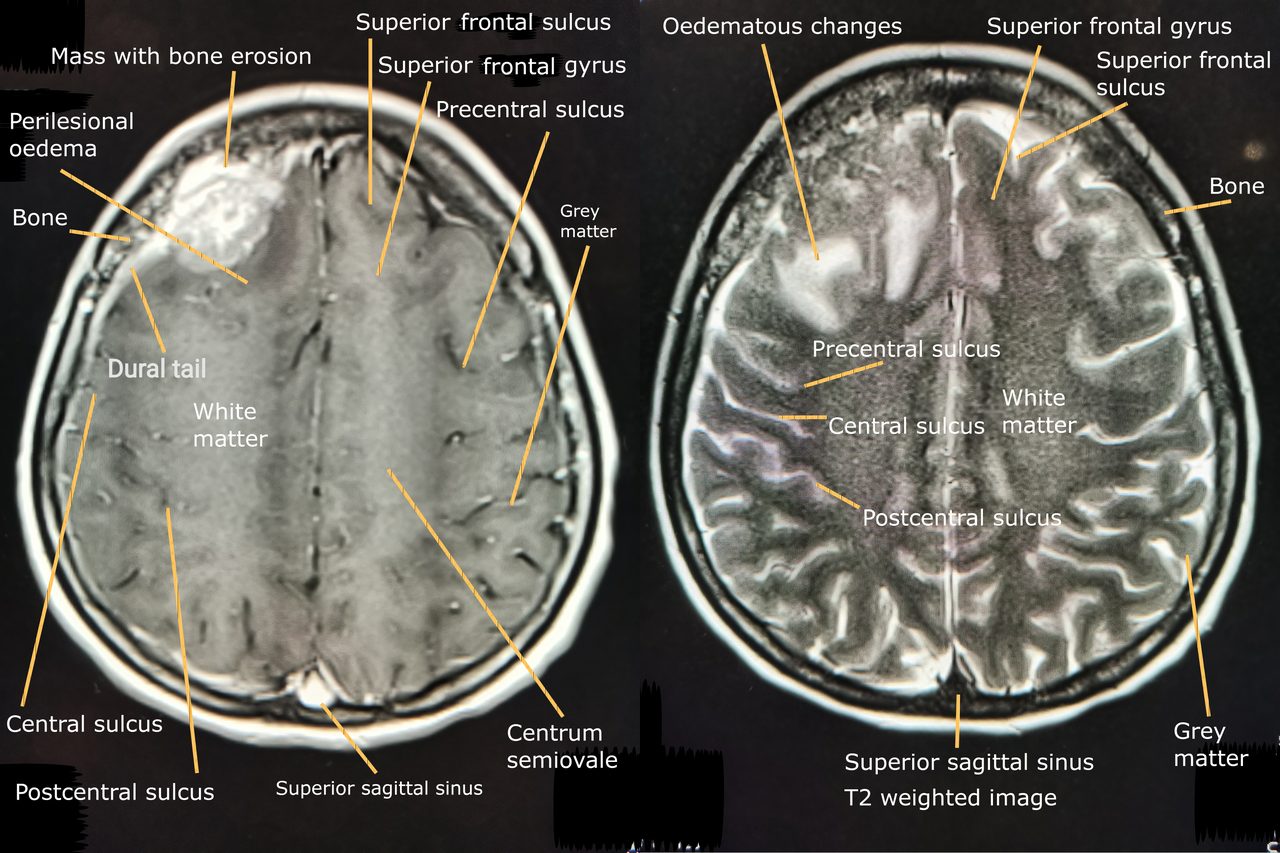
MRI Brain of extra-axial lesion at right frontal lobe with perilesional oedema. Left image: T1 weighted post gadolinium administration shows enhanced extra-axial mass with dural tail sign. Right image: T2 weighted image
Wikimedia Commons — File:MRI brain of extra-axial lesion at right frontal lobe with perilesional oedema.png | Cerevisae | CC BY-SA 4.0
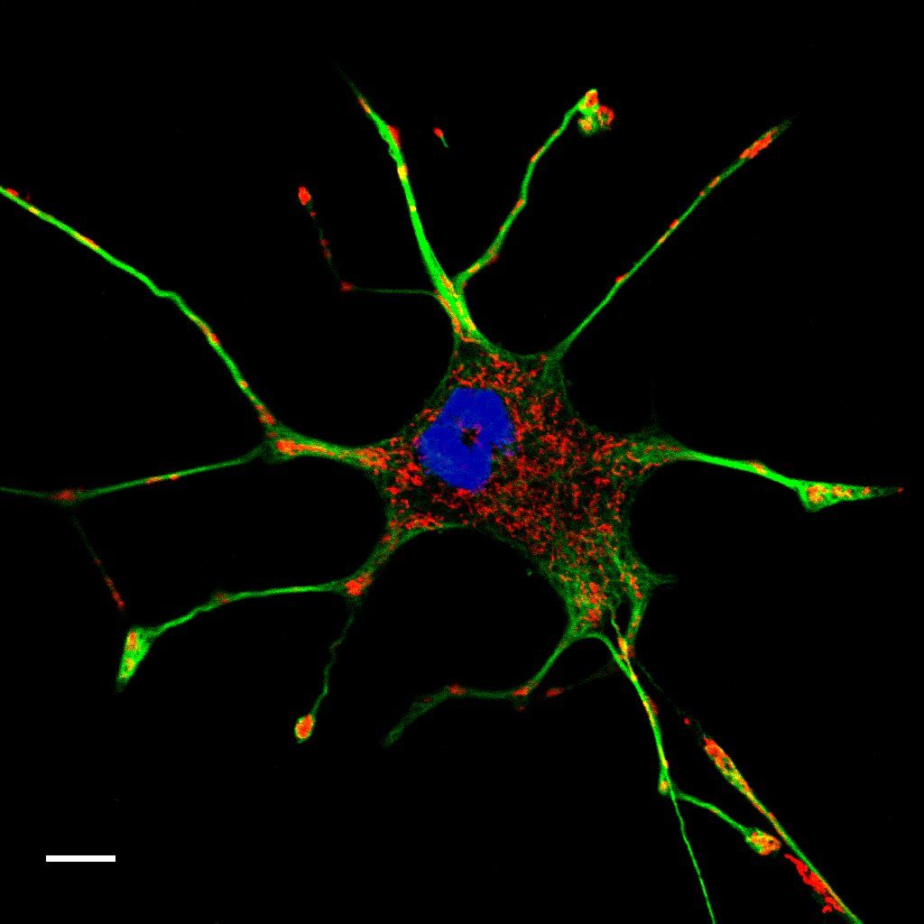
Mitochondria and cytoskeleton in PC-12 cells
Wikimedia Commons — File:Multicolor fluorescence image of a living PC-12 cell.jpeg | 8x57is | CC BY-SA 4.0
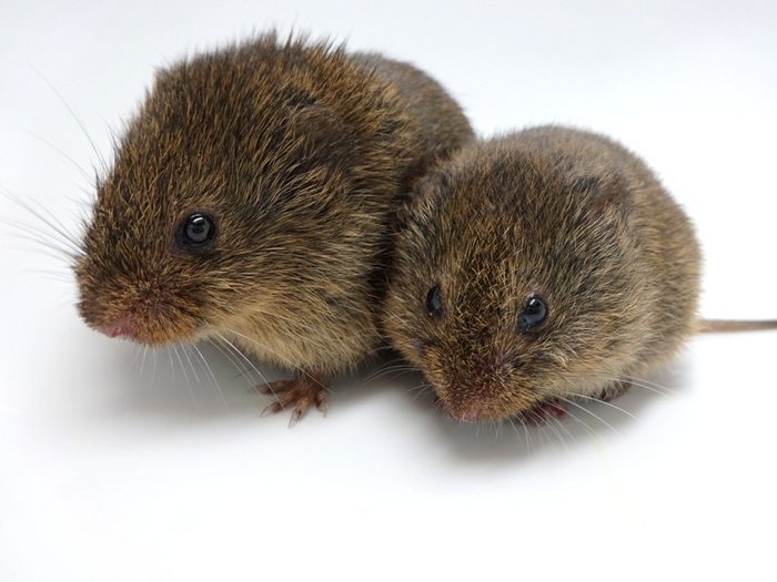
Prairie vole couple
Wikimedia Commons — File:Prairie Vole Nastacia Goodwin CC BY-SA.png | Nastacia Goodwin | CC BY-SA 4.0
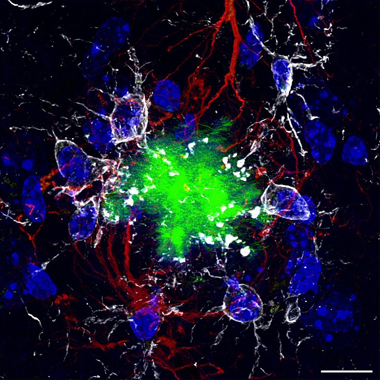
How can an explosion of colour be so damaging? How can so much light turn into absolute darkness? This is the sad reality that floods the brains of our Alzheimer's patients. The massive accumulation of these beta-amyloid
Wikimedia Commons — File:The Glowing Emptiness of an Alzheimer's Disease Patient.jpg | Nicocapelo | CC BY-SA 4.0
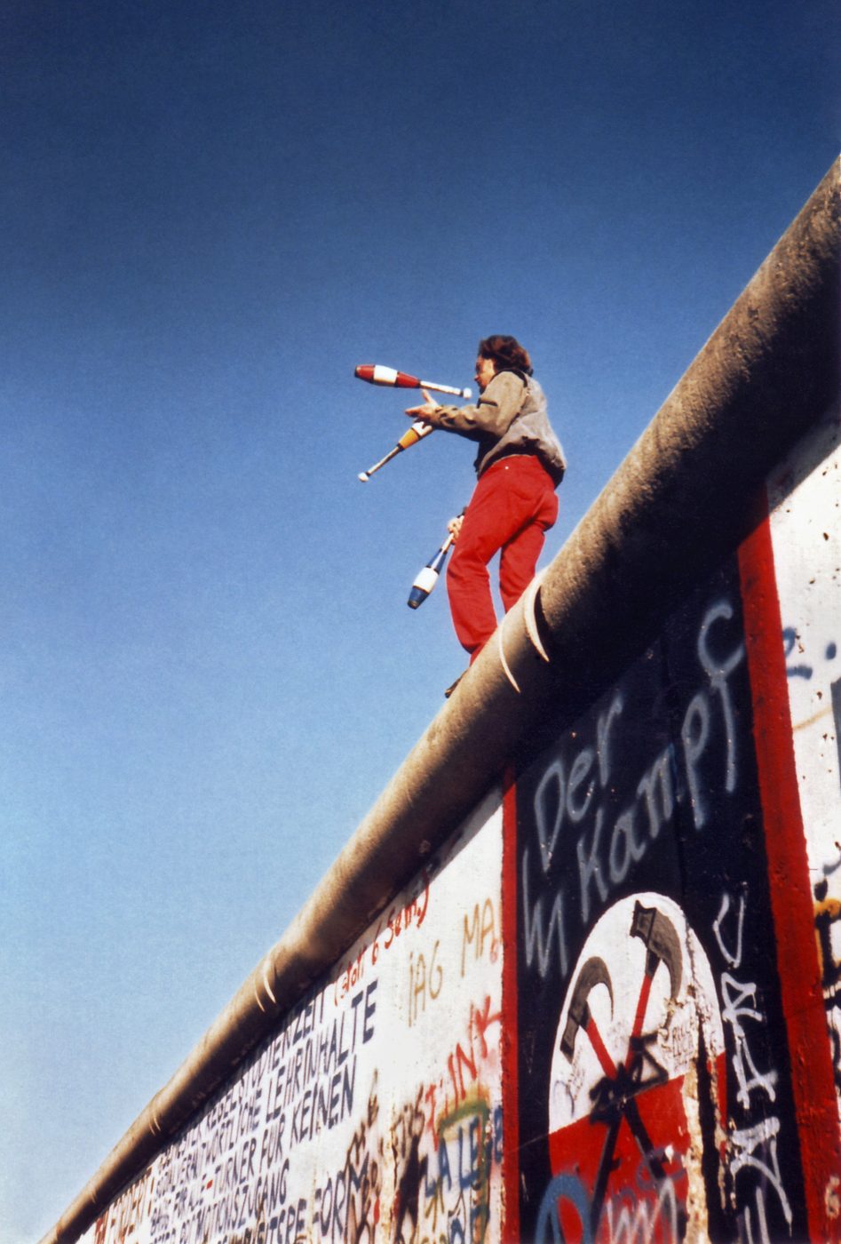
Juggling on the Berlin Wall on 16. November 1989. The black, white and red graffitti banner on the wall below the juggler depicts a pair of marching hammers, an allusion to the film Pink Floyd The Wall. The text sprayed
Wikimedia Commons — File:Juggling on the Berlin Wall 1a.jpg | Juggling_on_the_Berlin_Wall_1.jpg: Yann Forget derivative work: Durova (talk) | CC BY-SA 3.0
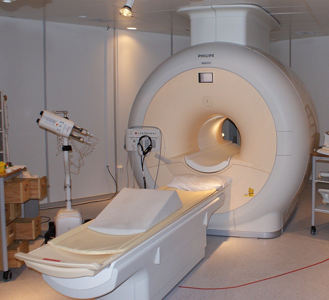
Philips MRI in Sahlgrenska Universitetsjukhuset, Gothenburg, Sweden
Wikimedia Commons — File:MRI-Philips.JPG | Jan Ainali | CC BY 3.0
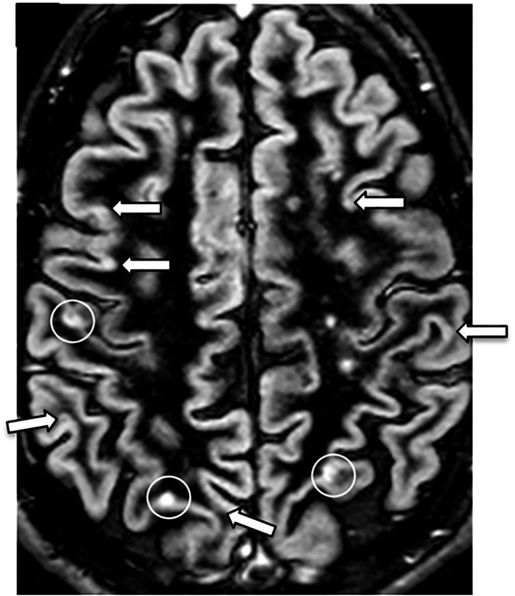
Double inversion recovery (DIR) MRI in the axial plane of the brain, showing lesions by multiple sclerosis. White circles surround two cortical lesions and one juxtacortical lesion. A separate PSIR MRI confirmed these, a
Wikimedia Commons — File:Axial DIR MRI of a brain with multiple sclerosis lesions.jpg | Article authors: Alice Favaretto, Davide Poggiali, Andrea Lazzarotto, Giuseppe Rolma, Francesco Causin, Paolo Gallo | CC BY 4.0
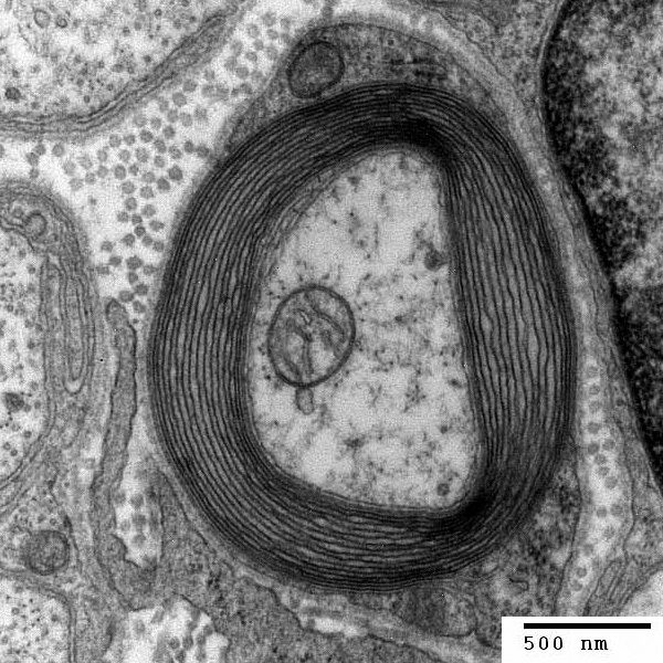
Transmission electron micrograph of a myelinated axon. The myelin layer (concentric) surrounds the axon of a neurone, showing cytoplasmathic organs inside. Generated and deposited into the public domain by the Electron M
Wikimedia Commons — File:Myelinated neuron.jpg | user:Roadnottaken | CC BY-SA 3.0
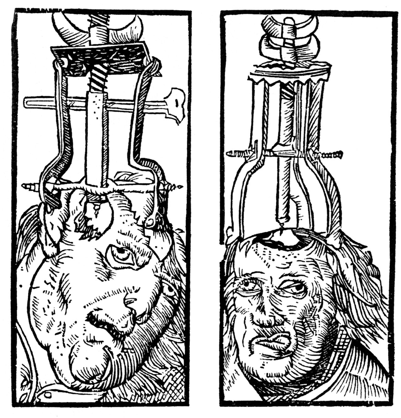
Engraving by Peter Treveris of a trepanation. From Heironymus von Braunschweig's Handywarke of surgeri
Wikimedia Commons — File:Peter Treveris - engraving of Trepanation for Handywarke of surgeri 1525.png | Peter Treveris | Public domain
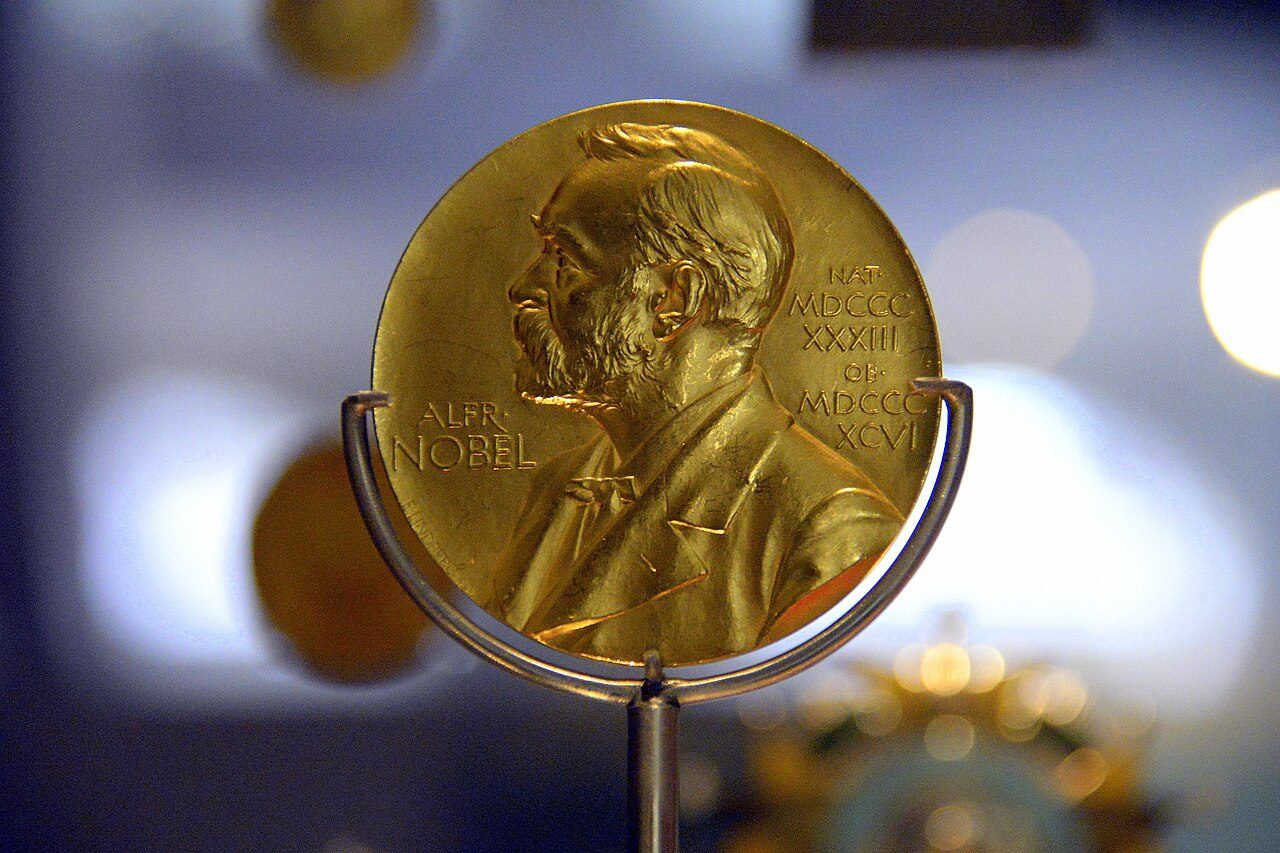
Nobel prize medal for medicine, Sweden, 1945, to Sir Alexander Fleming (1881-1955) who discovered Penicillin. On display at the National Museum of Scotland, Edinburgh, Scotland, UK
Wikimedia Commons — File:Nobel prize medal for medicine, Sweden, 1945, to Sir Alexander Fleming (1881-1955) who discovered Penicillin. On display at the National Museum of Scotland.jpg | Osama Shukir Muhammed Amin FRCP(Glasg) | CC BY-SA 4.0
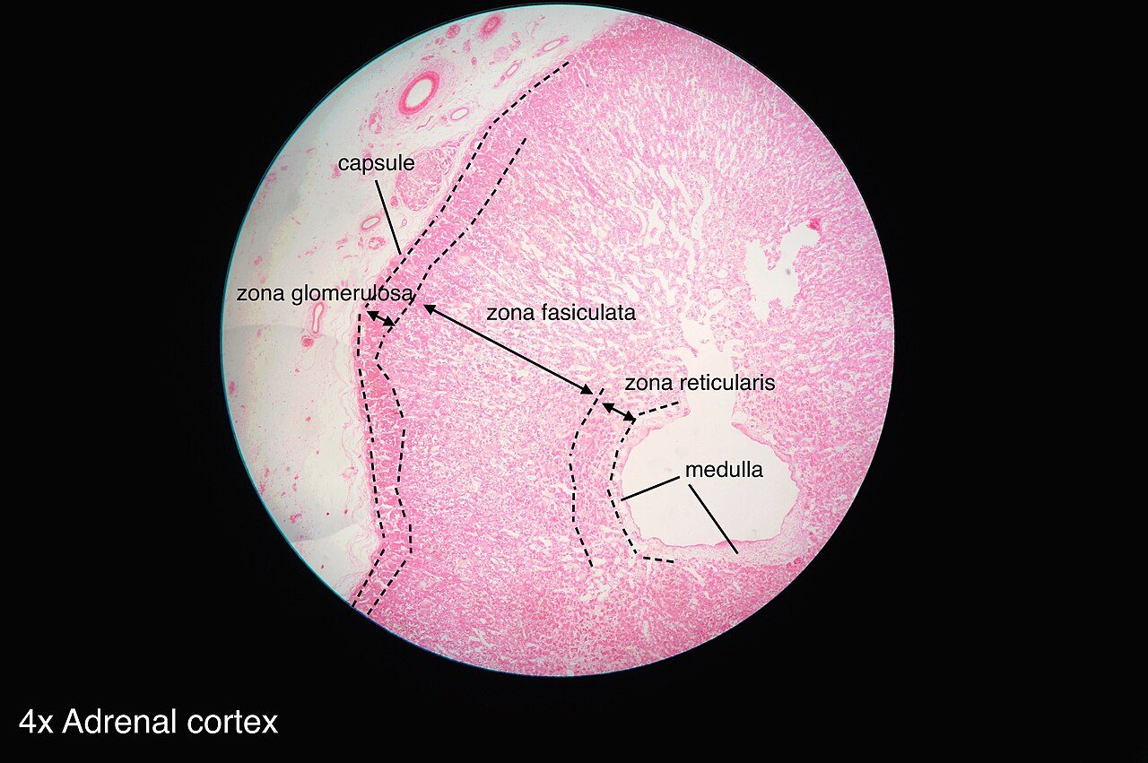
Histology of adrenal gland
Wikimedia Commons — File:Adrenal gland histology 2014.jpg | Athikhun.suw | CC BY-SA 4.0
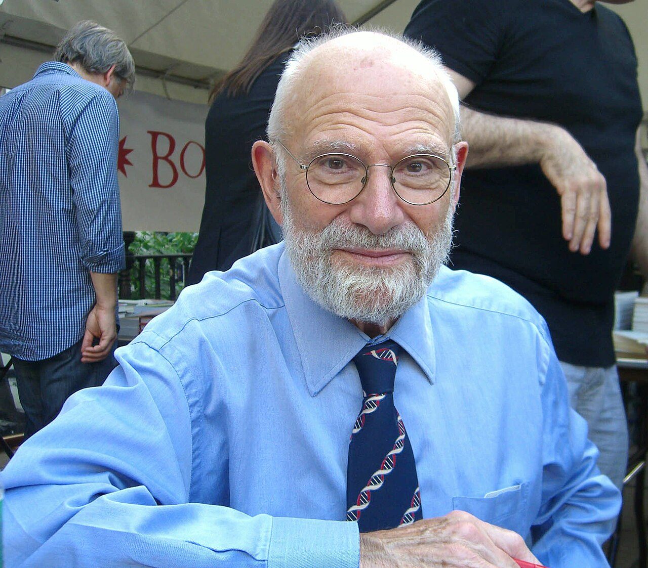
Neurologist and writer Oliver Sacks at the 2009 Brooklyn Book Festival, wearing a light-blue shirt and a dark-blue necktie that features a double helix pattern
Wikimedia Commons — File:9.13.09OliverSacksByLuigiNovi.jpg | Luigi Novi | CC BY 3.0
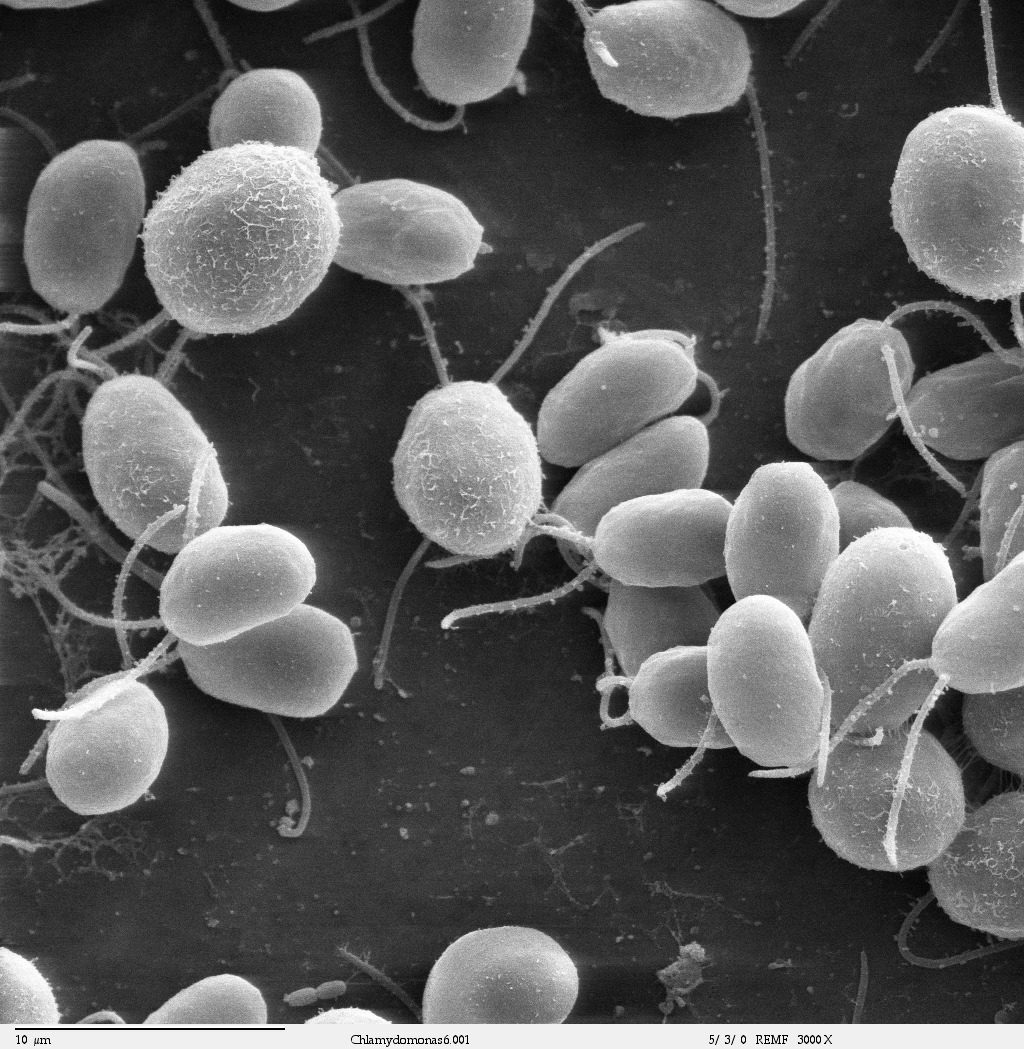
Scanning electron microscope image, showing an example of green algae (Chlorophyta). Chlamydomonas reinhardtii is a unicellular flagellate used as a model system in molecular genetics work and flagellar motility studies.
Wikimedia Commons — File:Chlamydomonas6-1.jpg | Dartmouth Electron Microscope Facility, Dartmouth College | Public domain
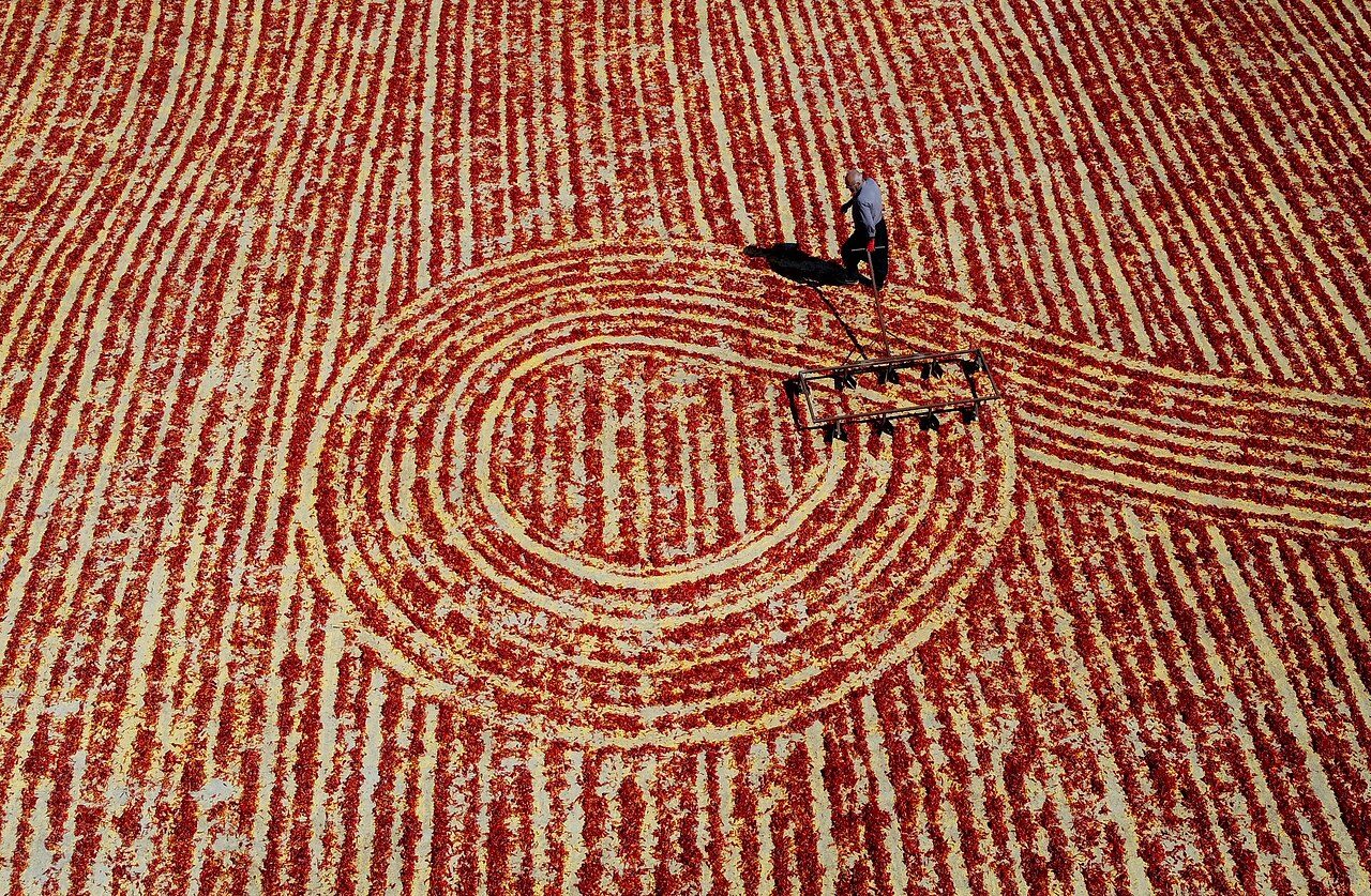
A man turns chili peppers laid out to dry using the traditional method in Turkey
Wikimedia Commons — File:Man turns drying chili peppers, Turkey.jpg | Rotadefterim | CC BY-SA 4.0
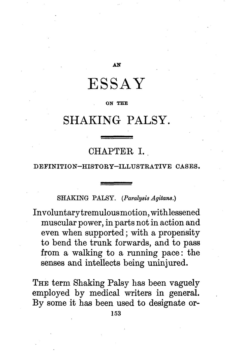
First page of James Parkinson's landmark 1817 work, An Essay on the Shaking Palsy, providing the first description of the disease that would bear his name
Wikimedia Commons — File:Parkinson, An Essay on the Shaking Palsy (first page).png | James Parkinson (1755–1824). Typeset and printed in 1817 by Whittingham & Rowland of London | Public domain
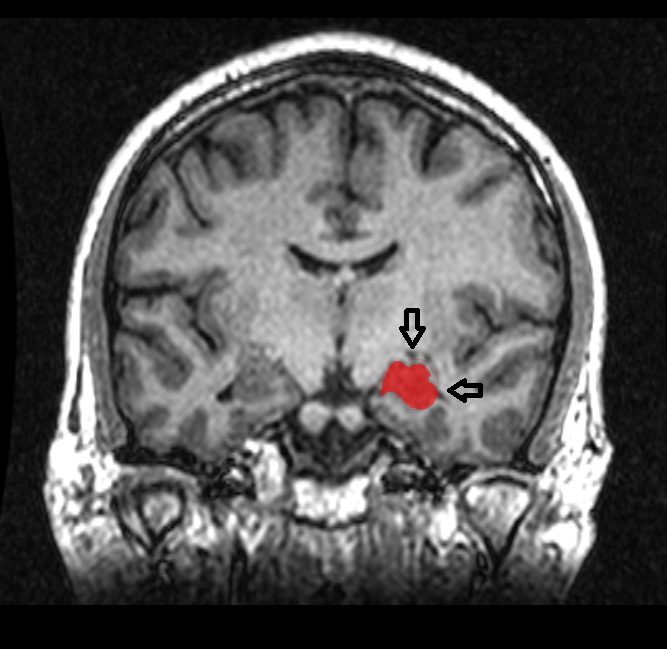
MRI coronal view of the amygdala
Wikimedia Commons — File:MRI Location Hippocampus up..png | Amber Rieder, Jenna Traynor | CC0
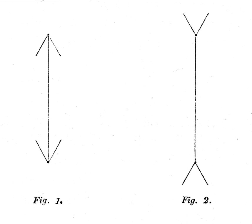
Müller-Lyer illusion - Franz Bretano. Optical illusion in which the orientation of arrowheads makes one line segment look longer than another. In the above figure, the line segments on the left and right are of equal len
Wikimedia Commons — File:Müller-Lyer illusion - Franz Bretano - 1892.png | Franz Carl Müller-Lyer, Franz Brentano | Public domain
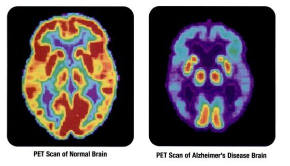
PET scans showing the differances between a normal older adult's brain and the brain of an older adult afflicted with Alzheimer's disease
Wikimedia Commons — File:PET scan-normal brain-alzheimers disease brain.PNG | Health and Human Services Department, National Institutes of Health, National Institute on Aging : p.24[1] | Public domain
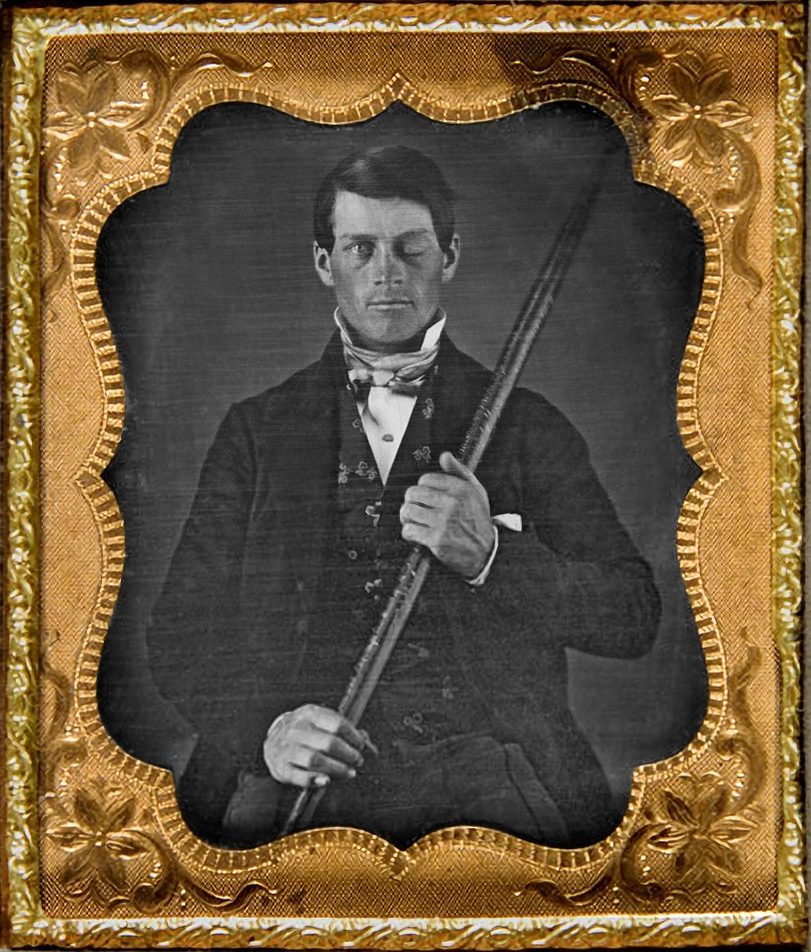
Photograph of cased-daguerreotype studio portrait of brain-injury survivor Phineas P. Gage (1823–1860) shown holding the tamping iron which injured him. Includes view of original embossed brass mat. From the collection o
Wikimedia Commons — File:Phineas Gage Cased Daguerreotype WilgusPhoto2008-12-19 EnhancedRetouched Color.jpg | Photograph by Jack and Beverly Wilgus of daguerreotype originally from their collection, and now in the Warren Anatomica | CC BY-SA 3.0
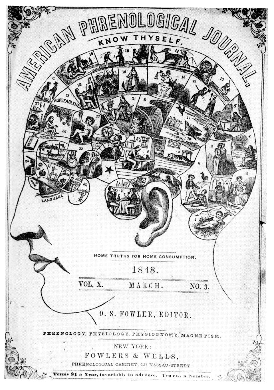
Cover of American Phrenological Journal from March 1848, volume 10, number 3
Wikimedia Commons — File:Phrenology journal (1848).jpg | Unknown | Public domain
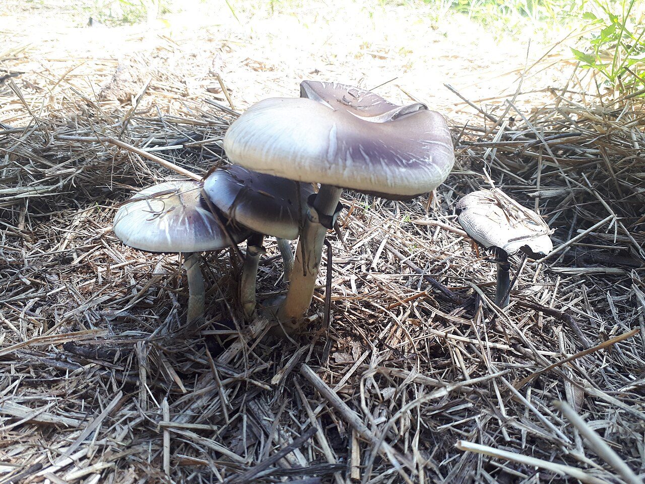
Psilocybe Cubensis Imire (Psilocybe Cubensis Zimbabwe)
Wikimedia Commons — File:Psilocybe Cubensis Imire (Psilocybe Cubensis Zimbabwe) 9.jpg | Robert van der Horst | CC BY-SA 4.0
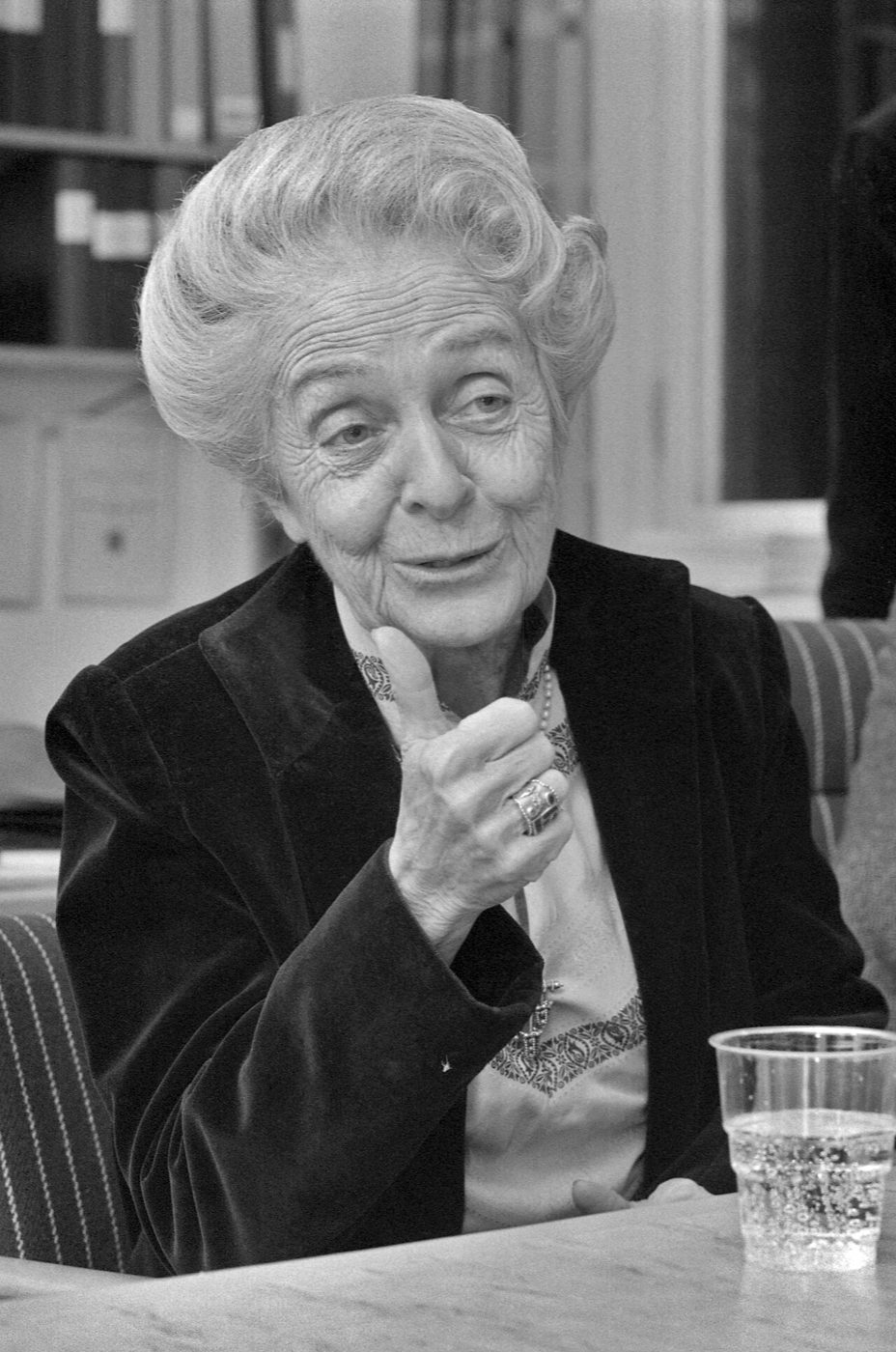
Rita Levi-Montalcini during a visit to Lund, Sweden, in 1986
Wikimedia Commons — File:Rita Levi-Montalcini (1986).jpg | Kurt Hagblom, Firma Hagblom-Foto, restored by Adam Cuerden | Public domain
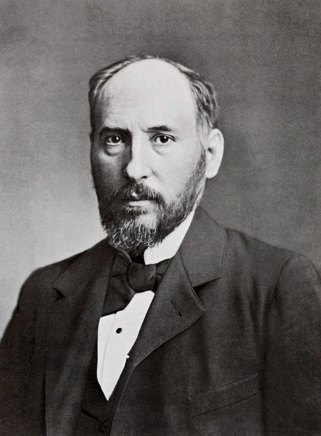
Santiago Ramón y Cajal. Spanish Nobel laureate in medicine. Restored jpg version of File:Cajal.PNG. Original image was a 5 Mb png but restoration was performed in tiff. File was mildly cropped. Dust and scratches were re
Wikimedia Commons — File:Santiago Ramón y Cajal (1852-1934) portrait (restored).jpg | Original photo is anonymous although published by Clark University in 1899. Restoration by Garrondo | Public domain
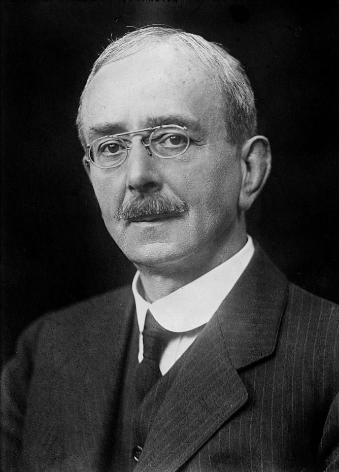
Prof. Scott Sherrington
Wikimedia Commons — File:Prof. Charles Scott Sherrington.jpg | No known restrictions on publication. Creator(s): Bain News Service, publisher | Public domain
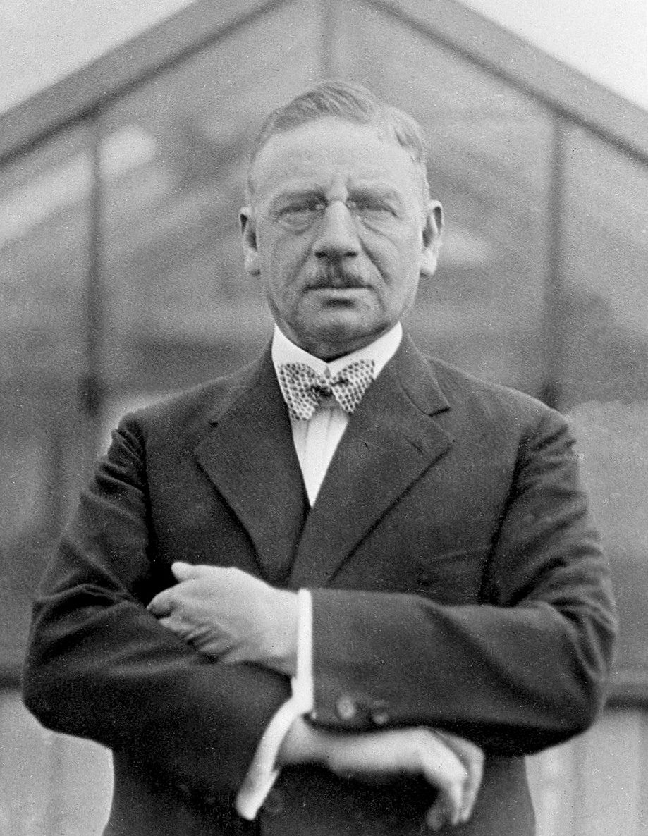
Otto Loewi; anon., at the International Physiological Congress in Boston
Wikimedia Commons — File:Otto Loewi 1929.jpg | Unknown authorUnknown author | CC BY 4.0
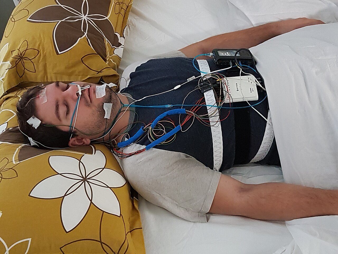
Posing with sleep testing equipment
Wikimedia Commons — File:Polysomnography model.jpg | Clinique Somnomed | CC BY-SA 4.0

Illustration of a busy office with employees rushing around and stressed with work. This is an image that is available for you to use as it has been licensed under the Attribution Creative Commons license. If you use it,
Wikimedia Commons — File:Workplace Stress Illustration.jpg | CIPHR Connect | CC BY 2.0
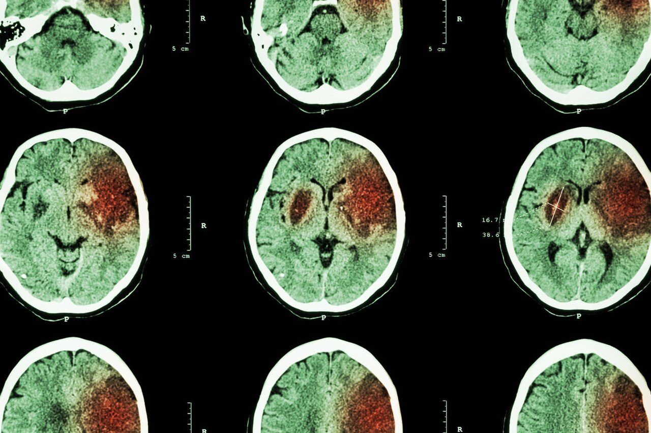
CT brain imaging used for rapid diagnosis of ischemic and hemorrhagic stroke
Wikimedia Commons — File:Brain CT Scan for Stroke Diagnosis.jpg | Goleisureintl | CC BY 4.0
Elderly woman walking towards the camera on a snowy walkway, Promenadeplatz, Munich, Germany
Wikimedia Commons — File:Elderly woman walking towards the camera on a snowy walkway, Promenadeplatz, Munich, Germany julesvernex2.jpg | Jules Verne Times Two | CC BY-SA 4.0
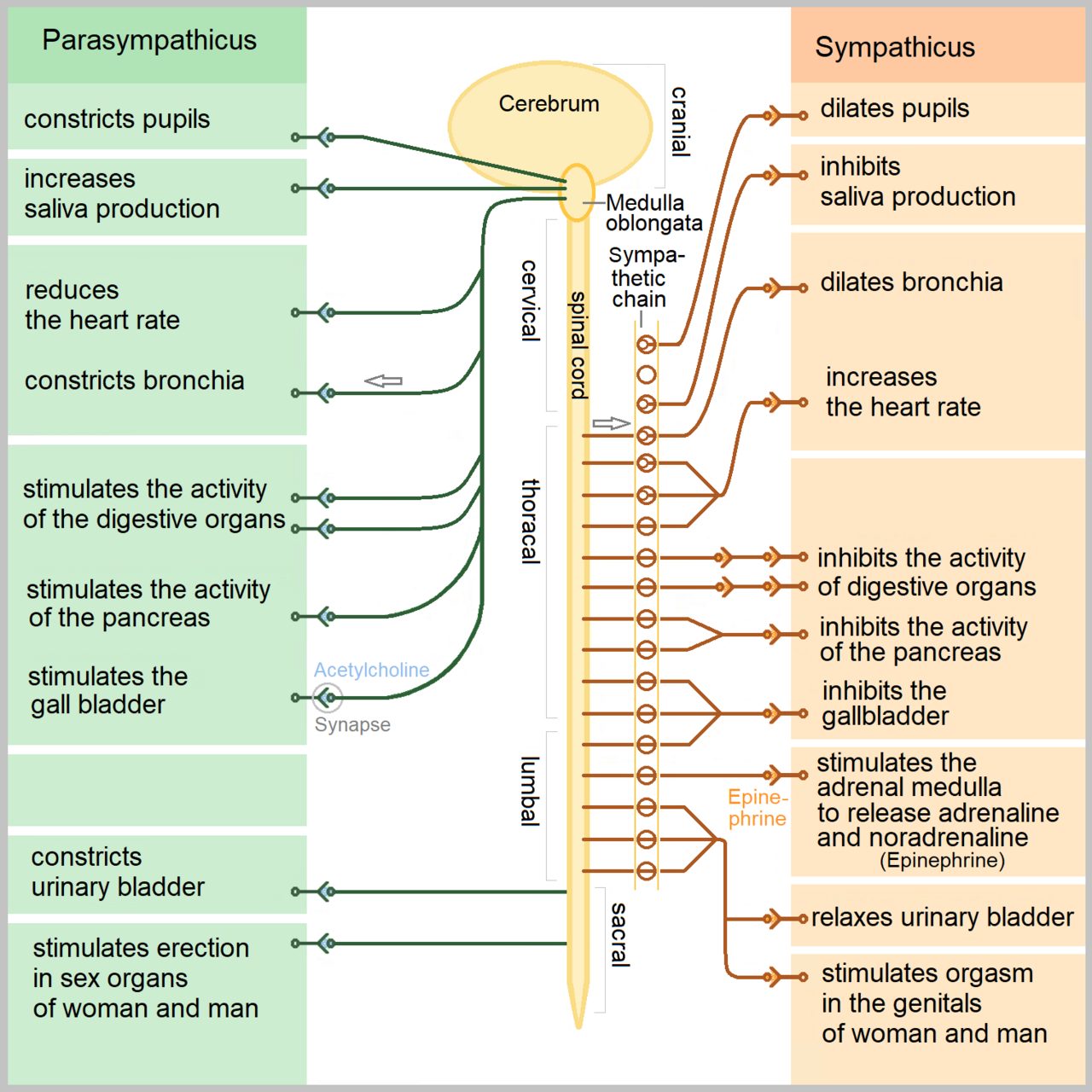
The human autonomic nervous system
Wikimedia Commons — File:The Autonomic Nervous System.png | Sciencia58 | CC0
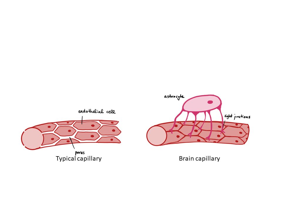
This figure illustrates the blood brain barrier, showing the difference between brain capillaries and capillaries in other tissues
Wikimedia Commons — File:Blood brain barrier.png | David Nascari and Alan Sved | CC BY-SA 4.0
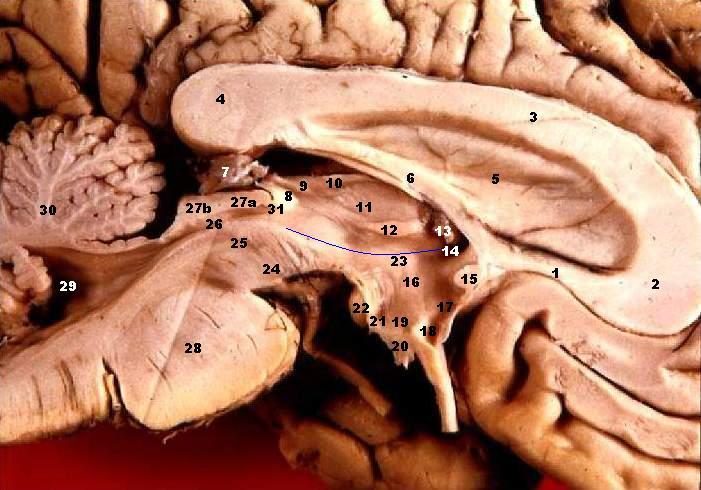
Human brain left - midsagitttal view - closeup Corpus callosum, Rostrum Corpus callosum, Genu Corpus callosum, Corpus Corpus callosum, Splenium Septum pellucidum Fornix, Corpus Glandula epiphysialis Recessus pinealis Hab
Wikimedia Commons — File:Human brain left midsagitttal view closeup description 2.JPG | John A Beal, PhD Dep't. of Cellular Biology & Anatomy, Louisiana State University Health Sciences Center Shreveport | CC BY 2.5
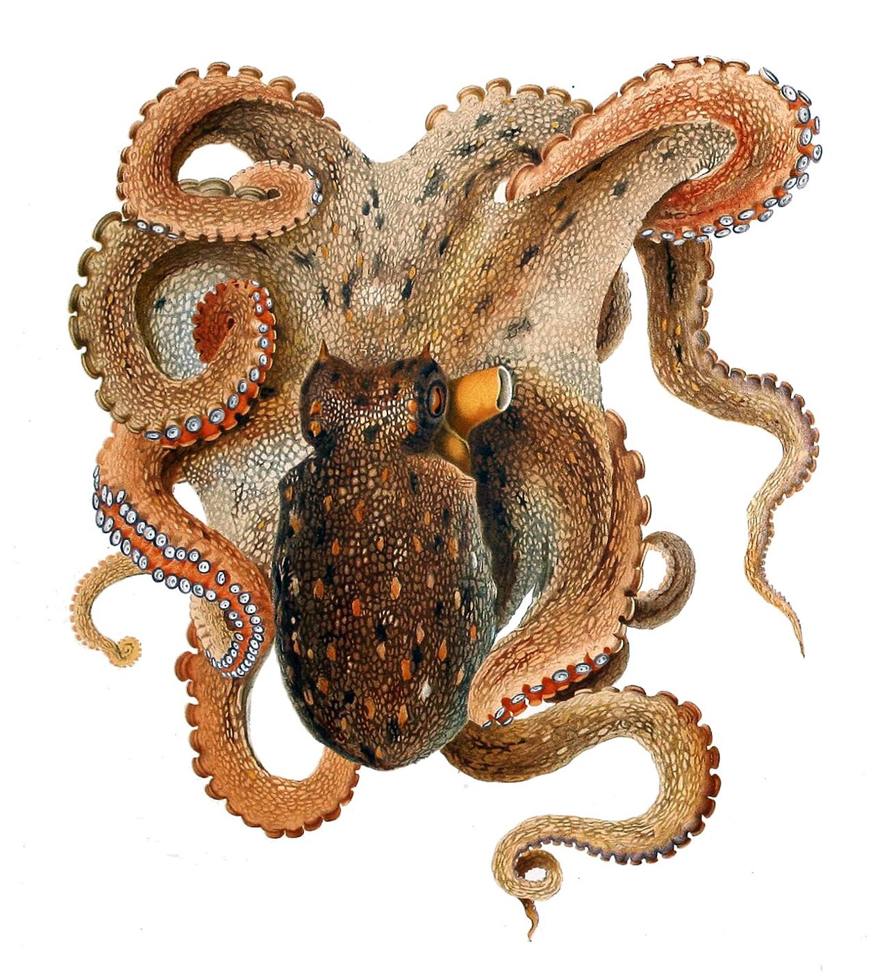
Octopus vulgaris
Wikimedia Commons — File:Octopus vulgaris Merculiano.jpg | Comingio Merculiano (1845-1915) in Jatta Giuseppe (1860-1903) | Public domain
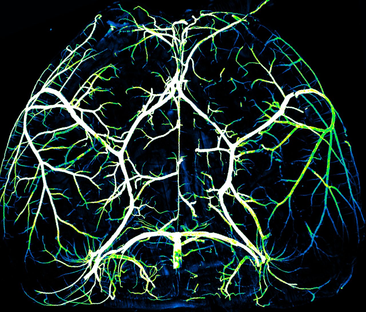
The arteries from the inner carotid arteries form the Circle of Willis, a structure that is conserved from mouse to humans
Wikimedia Commons — File:Circle of Willis.png | Brianwaichow | CC BY 4.0
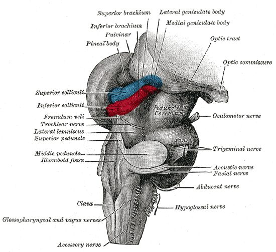
Hind- and mid-brains; postero-lateral view
Wikimedia Commons — File:Gray719.png | Henry Vandyke Carter | Public domain
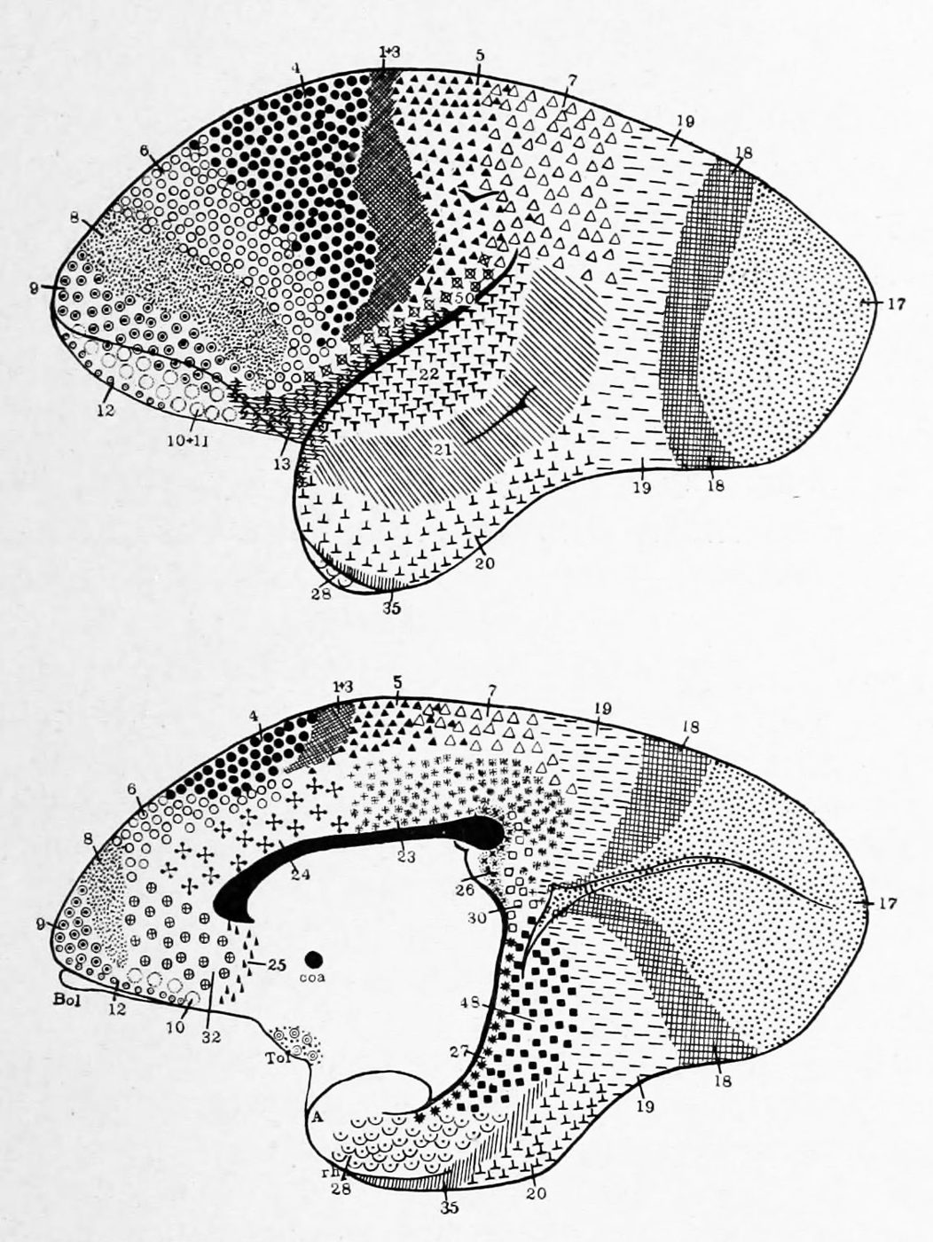
Brodmann areas of the marmoset (Hapale). 2:1. (K. Brodmann, 1909, p. 161, Fig. 96-97)
Wikimedia Commons — File:Brodmann areas in marmoset (K. Brodmann, 1909, p. 161, Fig. 96-97).jpg | Korbinian Brodmann | Public domain
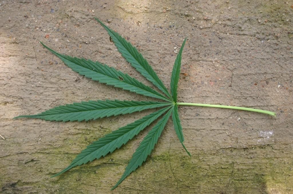
Image title: Cannabis sativa leaf Image from Public domain images website
Wikimedia Commons — File:Cannabis sativa leaf (1).jpg | Unknown authorUnknown author | Public domain
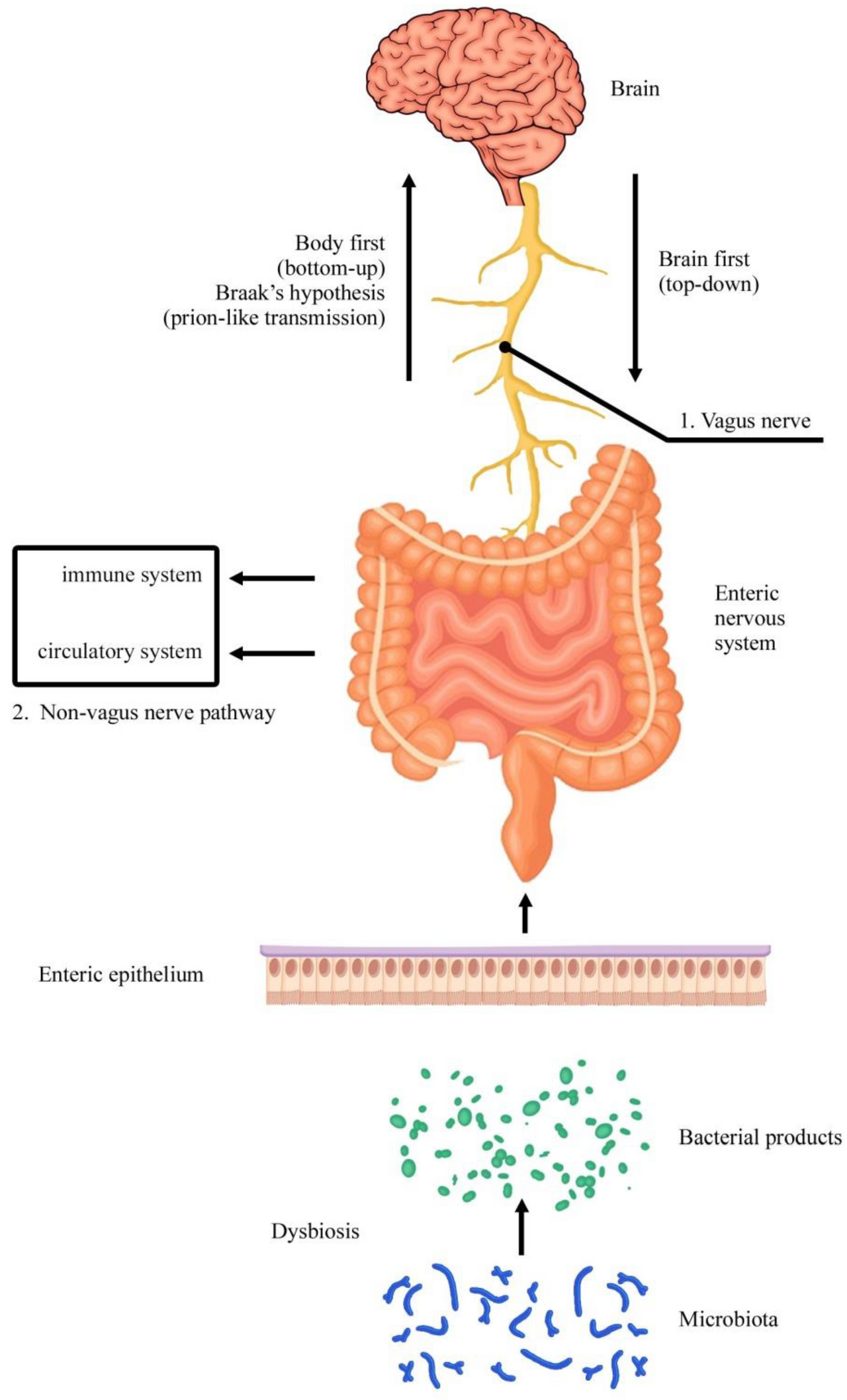
"Figure 1 Microbiota–gut–brain axis. Two subtypes of Parkinson’s disease patients have been recognized: a brain-first (top-down) type, where the hallmark of this pathology (α-synuclein) initially appears in the brain wit
Wikimedia Commons — File:Enteric Nervous System.png | Natale, Gianfranco, Larisa Ryskalin, Gabriele Morucci, Gloria Lazzeri, Alessandro Frati, and Francesco Fornai | CC BY 4.0
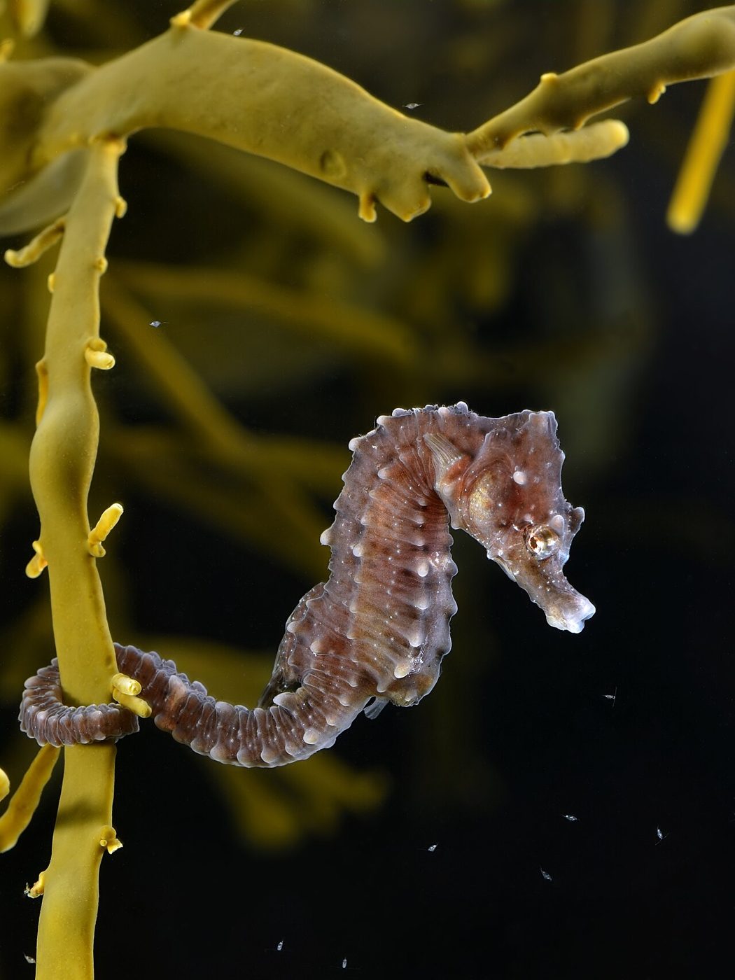
Short-snouted seahorse on knotted wrack. The animal was caught at 51°36′18.48″N 2°40′56.92″E / 51.6051333°N 2.6824778°E / 51.6051333; 2.6824778 on the Oosthinder banks in the Belgian part of the North Sea
Wikimedia Commons — File:Hippocampus hippocampus (on Ascophyllum nodosum).jpg | Hans Hillewaert | CC BY-SA 4.0
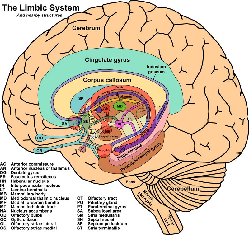
Cutout anatomy diagram of the human limbic system and some nearby structures
Wikimedia Commons — File:The Limbic System and Nearby Structures - John Taylor.jpg | John Taylor | Public domain

Norwegian prime minister Erna Solberg visits the Norwegian University of Science and Technology (NTNU), 4 March 2014. The Kavli Institute for Systems Neuroscience, Medical Technical Research Centre. Funding director of C
Wikimedia Commons — File:May-Britt Moser og Edvard Moser med statsminister Erna Solberg, NTNU 2014-03-04.jpg | Foto: Henrik Fjørtoft/NTNU Komm.avd | CC BY-SA 2.0
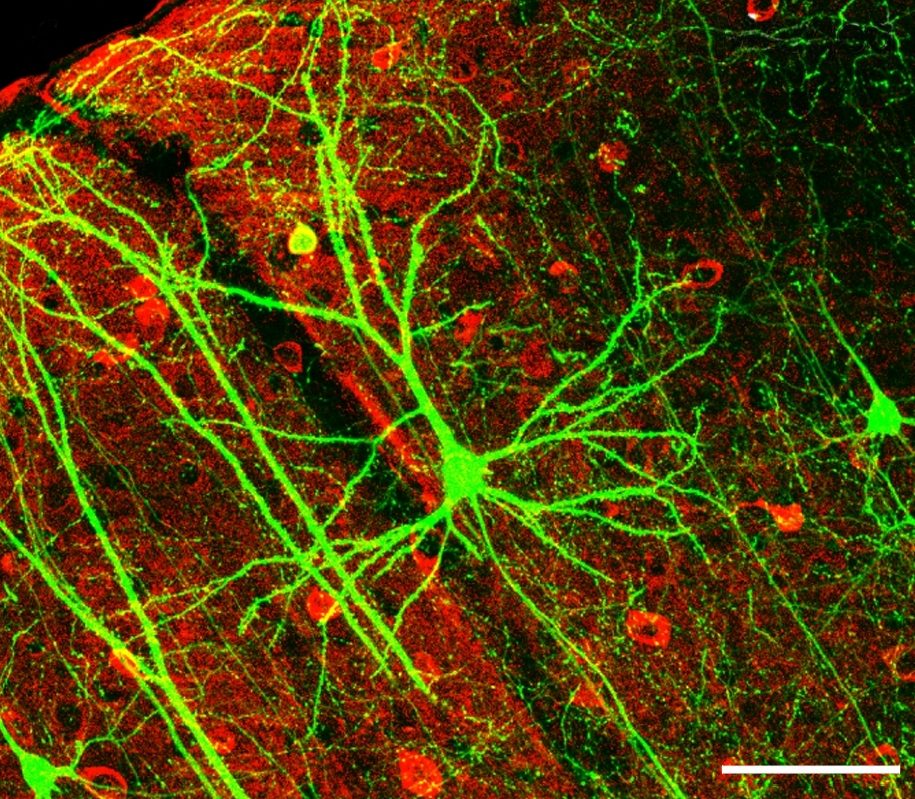
After the original figure legend: Coronal section containing the chronically imaged pyramidal neuron “dow” (visualized by green GFP) does not stain for GABA (visualized by antibody staining in red). Confocal image stack,
Wikimedia Commons — File:PLoSBiol4.e126.Fig6fNeuron.jpg | Wei-Chung Allen Lee, Hayden Huang, Guoping Feng, Joshua R. Sanes, Emery N. Brown, Peter T. So, Elly Nedivi | CC BY 2.5
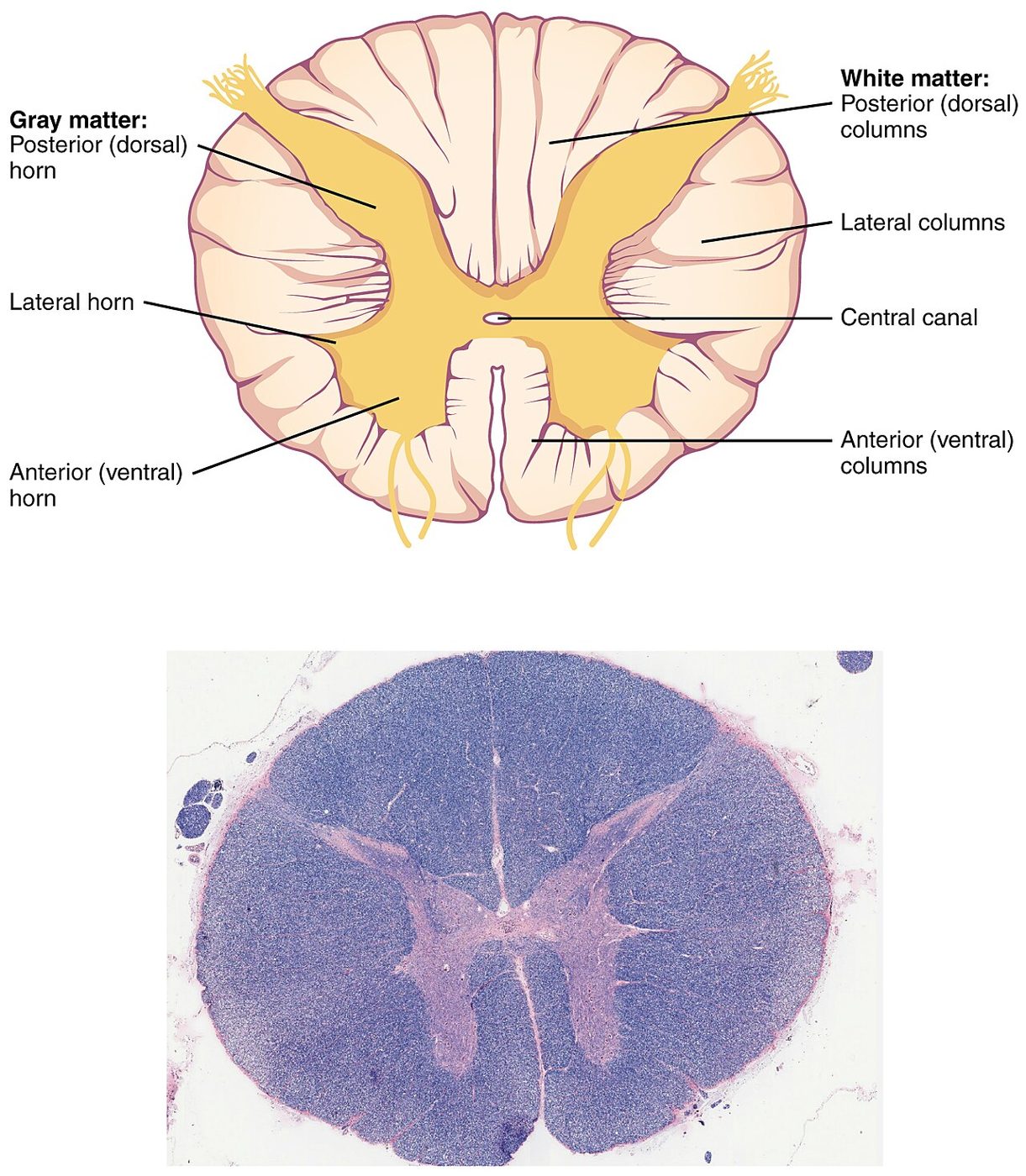
Version 8.25 from the Textbook OpenStax Anatomy and Physiology Published May 18, 2016
Wikimedia Commons — File:1313 Spinal Cord Cross Section.jpg | OpenStax | CC BY 4.0
Brailleskrift
Wikimedia Commons — File:Reading Braille.jpg | Stefan Malmesjö | CC BY 2.0
A Cornell University student created this album. Melvin Lorrel Nichols (1894-1981) was an undergrad from 1914-1918, and it is specifically those four years that his album meticulously depicts. It reveals an intimate and
Wikimedia Commons — File:This Unique Photo-Memory Album Offers a Fascinating Window to Student Life at Cornell University from 1914 to 1918.png | Stan S. Katz | CC BY-SA 4.0
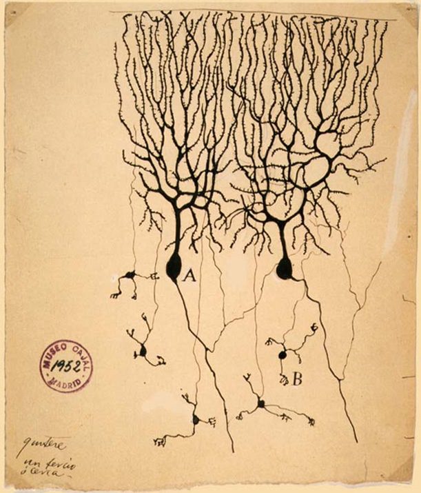
PurkinjeCell
Wikimedia Commons — File:PurkinjeCell.jpg | Unknown | Public domain
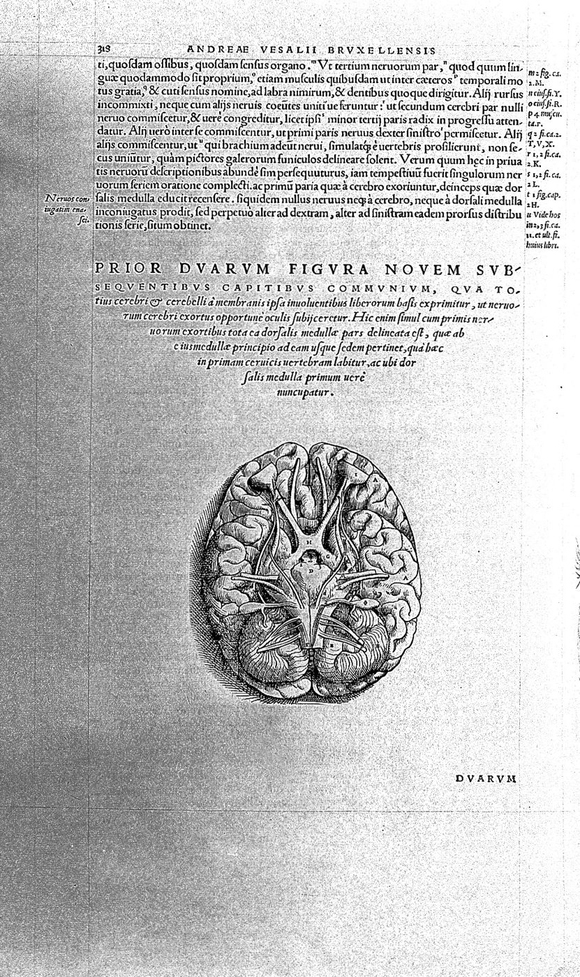
Cross section of the brain. Rare Books Keywords: Anatomy
Wikimedia Commons — File:Cross section of the brain, by Vesalius. Wellcome L0003676.jpg | Unknown | CC BY 4.0
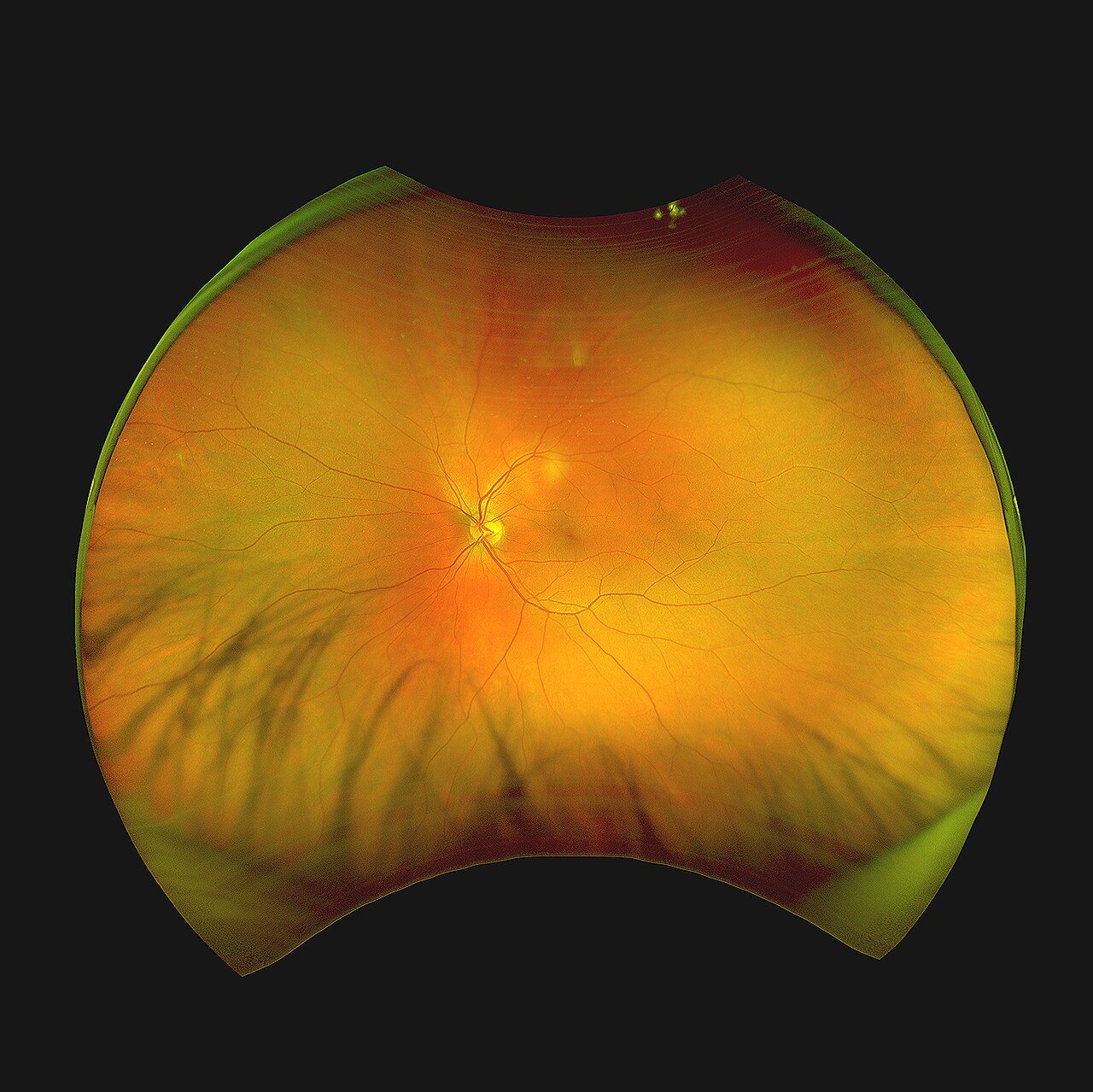
Fundus photograph of left retina of a 28 year old male
Wikimedia Commons — File:Fundus photograph Retina OS.jpg | Ktkvtsh | CC BY-SA 4.0
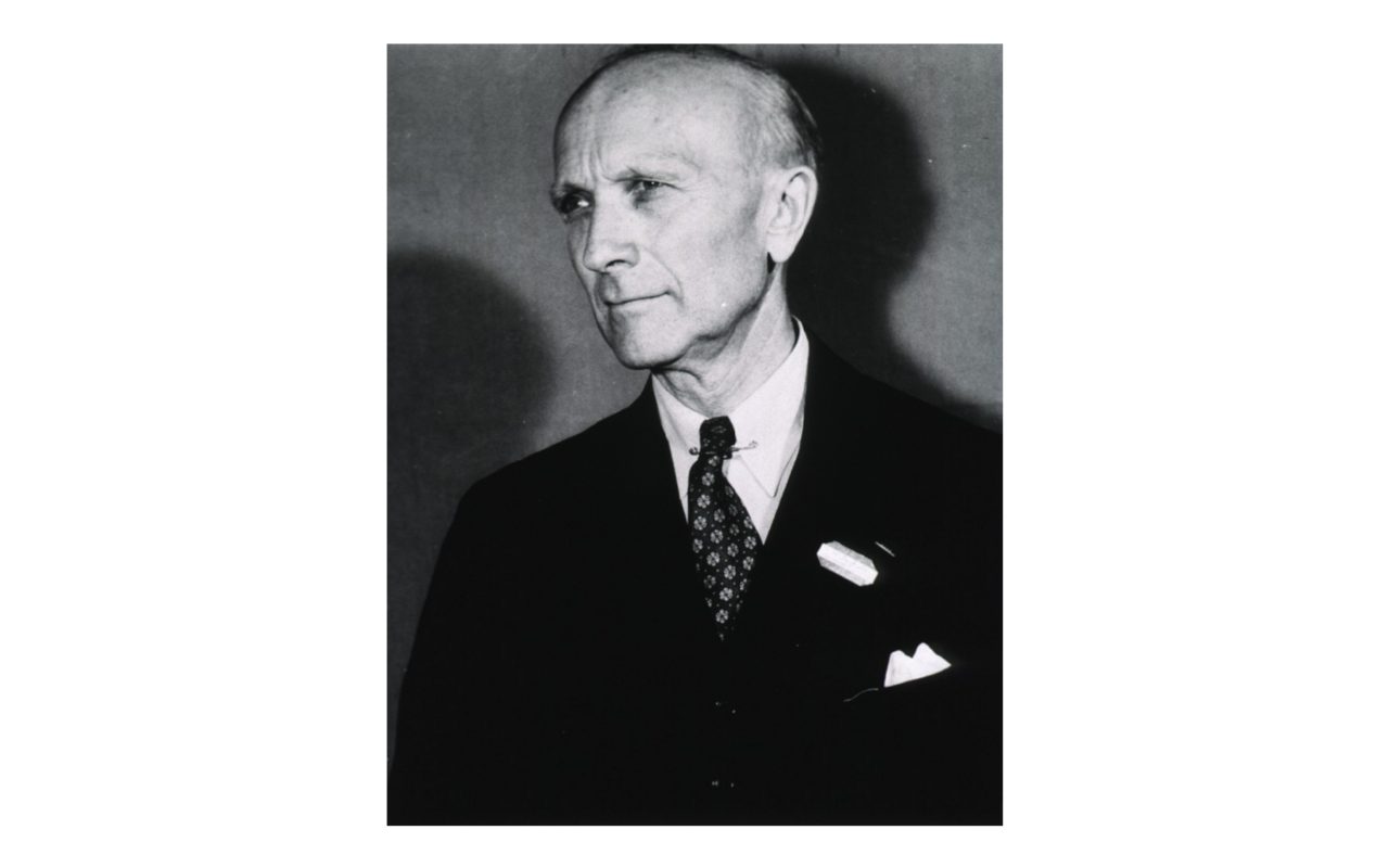
"Wilder G. Penfield". Collections of the National Library of Medicine. Portrait. Penfield, Wilder Graves 1891-1976. One photoprint reproduction. NLM Unique ID:101426095. Terms of Use: HMD provides access to digital image
Wikimedia Commons — File:Wilder Penfield.png | United States National Library of Medicine | Public domain
Original generative cover art for 'addiction'.
NeuroScience Decoded original artwork | NeuroScience Decoded | CC0 1.0
Original generative cover art for 'adhd'.
NeuroScience Decoded original artwork | NeuroScience Decoded | CC0 1.0
Original generative cover art for 'adult neurogenesis'.
NeuroScience Decoded original artwork | NeuroScience Decoded | CC0 1.0
Original generative cover art for 'als'.
NeuroScience Decoded original artwork | NeuroScience Decoded | CC0 1.0
Original generative cover art for 'antidepressants and psychiatric medication'.
NeuroScience Decoded original artwork | NeuroScience Decoded | CC0 1.0
Original generative cover art for 'anxiety disorders'.
NeuroScience Decoded original artwork | NeuroScience Decoded | CC0 1.0
Original generative cover art for 'astrocytes'.
NeuroScience Decoded original artwork | NeuroScience Decoded | CC0 1.0
Original generative cover art for 'attention'.
NeuroScience Decoded original artwork | NeuroScience Decoded | CC0 1.0
Original generative cover art for 'axon guidance and wiring'.
NeuroScience Decoded original artwork | NeuroScience Decoded | CC0 1.0
Original generative cover art for 'balance and proprioception'.
NeuroScience Decoded original artwork | NeuroScience Decoded | CC0 1.0
Original generative cover art for 'bipolar disorder'.
NeuroScience Decoded original artwork | NeuroScience Decoded | CC0 1.0
Original generative cover art for 'brain networks and the connectome'.
NeuroScience Decoded original artwork | NeuroScience Decoded | CC0 1.0
Original generative cover art for 'computational neuroscience'.
NeuroScience Decoded original artwork | NeuroScience Decoded | CC0 1.0

Original generative cover art for 'concussion tbi and cte'.
NeuroScience Decoded original artwork | NeuroScience Decoded | CC0 1.0
Original generative cover art for 'decision making'.
NeuroScience Decoded original artwork | NeuroScience Decoded | CC0 1.0
Original generative cover art for 'depression'.
NeuroScience Decoded original artwork | NeuroScience Decoded | CC0 1.0
Original generative cover art for 'descartes and the mechanical reflex'.
NeuroScience Decoded original artwork | NeuroScience Decoded | CC0 1.0
Original generative cover art for 'dreams'.
NeuroScience Decoded original artwork | NeuroScience Decoded | CC0 1.0
Original generative cover art for 'electrical synapses and gap junctions'.
NeuroScience Decoded original artwork | NeuroScience Decoded | CC0 1.0
Original generative cover art for 'endorphins and neuropeptides'.
NeuroScience Decoded original artwork | NeuroScience Decoded | CC0 1.0
Original generative cover art for 'eric kandel'.
NeuroScience Decoded original artwork | NeuroScience Decoded | CC0 1.0
Original generative cover art for 'executive function and the prefrontal cortex'.
NeuroScience Decoded original artwork | NeuroScience Decoded | CC0 1.0
Original generative cover art for 'fear and anxiety circuits'.
NeuroScience Decoded original artwork | NeuroScience Decoded | CC0 1.0
Original generative cover art for 'forgetting and false memories'.
NeuroScience Decoded original artwork | NeuroScience Decoded | CC0 1.0

Original generative cover art for 'free will and the brain'.
NeuroScience Decoded original artwork | NeuroScience Decoded | CC0 1.0
Original generative cover art for 'gene and cell therapies for the brain'.
NeuroScience Decoded original artwork | NeuroScience Decoded | CC0 1.0
Original generative cover art for 'glial cells'.
NeuroScience Decoded original artwork | NeuroScience Decoded | CC0 1.0

Original generative cover art for 'glutamate and gaba'.
NeuroScience Decoded original artwork | NeuroScience Decoded | CC0 1.0
Original generative cover art for 'golgi cajal and the neuron doctrine'.
NeuroScience Decoded original artwork | NeuroScience Decoded | CC0 1.0
Original generative cover art for 'hearing'.
NeuroScience Decoded original artwork | NeuroScience Decoded | CC0 1.0
Original generative cover art for 'hero'.
NeuroScience Decoded original artwork | NeuroScience Decoded | CC0 1.0

Original generative cover art for 'hormones and the brain'.
NeuroScience Decoded original artwork | NeuroScience Decoded | CC0 1.0
Original generative cover art for 'how brain drugs work'.
NeuroScience Decoded original artwork | NeuroScience Decoded | CC0 1.0
Original generative cover art for 'how memories are made ltp'.
NeuroScience Decoded original artwork | NeuroScience Decoded | CC0 1.0
Original generative cover art for 'how neurons communicate'.
NeuroScience Decoded original artwork | NeuroScience Decoded | CC0 1.0
Original generative cover art for 'how the brain builds itself'.
NeuroScience Decoded original artwork | NeuroScience Decoded | CC0 1.0

Original generative cover art for 'how the brain makes emotions'.
NeuroScience Decoded original artwork | NeuroScience Decoded | CC0 1.0

Original generative cover art for 'how the senses work'.
NeuroScience Decoded original artwork | NeuroScience Decoded | CC0 1.0
Original generative cover art for 'how we study the brain'.
NeuroScience Decoded original artwork | NeuroScience Decoded | CC0 1.0
Original generative cover art for 'hubel wiesel and the visual cortex'.
NeuroScience Decoded original artwork | NeuroScience Decoded | CC0 1.0

Original generative cover art for 'hunger thirst and appetite'.
NeuroScience Decoded original artwork | NeuroScience Decoded | CC0 1.0
Original generative cover art for 'intelligence and the brain'.
NeuroScience Decoded original artwork | NeuroScience Decoded | CC0 1.0
Original generative cover art for 'interoception'.
NeuroScience Decoded original artwork | NeuroScience Decoded | CC0 1.0
Original generative cover art for 'medieval and islamic brain science'.
NeuroScience Decoded original artwork | NeuroScience Decoded | CC0 1.0
Original generative cover art for 'memory consolidation'.
NeuroScience Decoded original artwork | NeuroScience Decoded | CC0 1.0
Original generative cover art for 'microscopy staining and clearing'.
NeuroScience Decoded original artwork | NeuroScience Decoded | CC0 1.0
Original generative cover art for 'migraine and headache'.
NeuroScience Decoded original artwork | NeuroScience Decoded | CC0 1.0
Original generative cover art for 'mirror neurons'.
NeuroScience Decoded original artwork | NeuroScience Decoded | CC0 1.0
Original generative cover art for 'neural coding'.
NeuroScience Decoded original artwork | NeuroScience Decoded | CC0 1.0

Original generative cover art for 'neuroethics'.
NeuroScience Decoded original artwork | NeuroScience Decoded | CC0 1.0
Original generative cover art for 'neurogenesis and neural migration'.
NeuroScience Decoded original artwork | NeuroScience Decoded | CC0 1.0
Original generative cover art for 'neuromorphic computing'.
NeuroScience Decoded original artwork | NeuroScience Decoded | CC0 1.0
Original generative cover art for 'neuromyths in education'.
NeuroScience Decoded original artwork | NeuroScience Decoded | CC0 1.0
Original generative cover art for 'neuroplasticity'.
NeuroScience Decoded original artwork | NeuroScience Decoded | CC0 1.0
Original generative cover art for 'neurorehabilitation'.
NeuroScience Decoded original artwork | NeuroScience Decoded | CC0 1.0
Original generative cover art for 'neuroscience and the law'.
NeuroScience Decoded original artwork | NeuroScience Decoded | CC0 1.0
Original generative cover art for 'neurotransmitter release'.
NeuroScience Decoded original artwork | NeuroScience Decoded | CC0 1.0
Original generative cover art for 'neurotransmitters overview'.
NeuroScience Decoded original artwork | NeuroScience Decoded | CC0 1.0
Original generative cover art for 'ocd'.
NeuroScience Decoded original artwork | NeuroScience Decoded | CC0 1.0
Original generative cover art for 'place cells grid cells and navigation'.
NeuroScience Decoded original artwork | NeuroScience Decoded | CC0 1.0
Original generative cover art for 'ptsd'.
NeuroScience Decoded original artwork | NeuroScience Decoded | CC0 1.0
Original generative cover art for 'receptors ionotropic and metabotropic'.
NeuroScience Decoded original artwork | NeuroScience Decoded | CC0 1.0
Original generative cover art for 'recording single neurons and patch clamp'.
NeuroScience Decoded original artwork | NeuroScience Decoded | CC0 1.0
Original generative cover art for 'reflexes'.
NeuroScience Decoded original artwork | NeuroScience Decoded | CC0 1.0
Original generative cover art for 'saltatory conduction'.
NeuroScience Decoded original artwork | NeuroScience Decoded | CC0 1.0
Original generative cover art for 'schizophrenia'.
NeuroScience Decoded original artwork | NeuroScience Decoded | CC0 1.0

Original generative cover art for 'serotonin'.
NeuroScience Decoded original artwork | NeuroScience Decoded | CC0 1.0

Original generative cover art for 'smell and taste'.
NeuroScience Decoded original artwork | NeuroScience Decoded | CC0 1.0
Original generative cover art for 'synaptic integration'.
NeuroScience Decoded original artwork | NeuroScience Decoded | CC0 1.0
Original generative cover art for 'synaptic pruning'.
NeuroScience Decoded original artwork | NeuroScience Decoded | CC0 1.0

Original generative cover art for 'the action potential'.
NeuroScience Decoded original artwork | NeuroScience Decoded | CC0 1.0
Original generative cover art for 'the amygdala'.
NeuroScience Decoded original artwork | NeuroScience Decoded | CC0 1.0
Original generative cover art for 'the basal ganglia'.
NeuroScience Decoded original artwork | NeuroScience Decoded | CC0 1.0
Original generative cover art for 'the birth of modern neuroscience'.
NeuroScience Decoded original artwork | NeuroScience Decoded | CC0 1.0
Original generative cover art for 'the brain and mental illness'.
NeuroScience Decoded original artwork | NeuroScience Decoded | CC0 1.0
Original generative cover art for 'the brain vs artificial neural networks'.
NeuroScience Decoded original artwork | NeuroScience Decoded | CC0 1.0
Original generative cover art for 'the cerebellum'.
NeuroScience Decoded original artwork | NeuroScience Decoded | CC0 1.0
Original generative cover art for 'the frontal lobe'.
NeuroScience Decoded original artwork | NeuroScience Decoded | CC0 1.0
Original generative cover art for 'the future of neuroscience'.
NeuroScience Decoded original artwork | NeuroScience Decoded | CC0 1.0
Original generative cover art for 'the glymphatic system'.
NeuroScience Decoded original artwork | NeuroScience Decoded | CC0 1.0
Original generative cover art for 'the hypothalamus and pituitary'.
NeuroScience Decoded original artwork | NeuroScience Decoded | CC0 1.0
Original generative cover art for 'the motor cortex and homunculus'.
NeuroScience Decoded original artwork | NeuroScience Decoded | CC0 1.0
Original generative cover art for 'the nervous system at a glance'.
NeuroScience Decoded original artwork | NeuroScience Decoded | CC0 1.0
Original generative cover art for 'the occipital lobe'.
NeuroScience Decoded original artwork | NeuroScience Decoded | CC0 1.0
Original generative cover art for 'the parietal lobe'.
NeuroScience Decoded original artwork | NeuroScience Decoded | CC0 1.0
Original generative cover art for 'the peripheral nervous system'.
NeuroScience Decoded original artwork | NeuroScience Decoded | CC0 1.0
Original generative cover art for 'the predictive brain'.
NeuroScience Decoded original artwork | NeuroScience Decoded | CC0 1.0

Original generative cover art for 'the resting membrane potential'.
NeuroScience Decoded original artwork | NeuroScience Decoded | CC0 1.0

Original generative cover art for 'the reward system'.
NeuroScience Decoded original artwork | NeuroScience Decoded | CC0 1.0
Original generative cover art for 'the social brain'.
NeuroScience Decoded original artwork | NeuroScience Decoded | CC0 1.0
Original generative cover art for 'the synapse'.
NeuroScience Decoded original artwork | NeuroScience Decoded | CC0 1.0
Original generative cover art for 'the teenage brain'.
NeuroScience Decoded original artwork | NeuroScience Decoded | CC0 1.0
Original generative cover art for 'the temporal lobe'.
NeuroScience Decoded original artwork | NeuroScience Decoded | CC0 1.0
Original generative cover art for 'the thalamus'.
NeuroScience Decoded original artwork | NeuroScience Decoded | CC0 1.0
Original generative cover art for 'theories of consciousness'.
NeuroScience Decoded original artwork | NeuroScience Decoded | CC0 1.0
Original generative cover art for 'what is consciousness'.
NeuroScience Decoded original artwork | NeuroScience Decoded | CC0 1.0
Original generative cover art for 'what is neuroscience'.
NeuroScience Decoded original artwork | NeuroScience Decoded | CC0 1.0
Original generative cover art for 'why we sleep'.
NeuroScience Decoded original artwork | NeuroScience Decoded | CC0 1.0
Notes
NeuroScience Decoded uses 86 photographs and historical images from Wikimedia Commons alongside original teaching diagrams and generative cover art made for this site.
Every Wikimedia Commons image used here was downloaded, downscaled and re-hosted on this site — nothing is hot-linked. Each is public domain or carries a Creative Commons licence (CC0, CC BY or CC BY-SA); its Commons file name, author and licence are listed above as the licence requires.
The teaching diagrams and the generative cover plates were made for this site and are released under CC0 (public domain dedication).
This site contains no links to other websites and loads nothing from outside servers: every image, font and script is stored locally. For further study, each article suggests plain-text phrases you can type into YouTube's search box yourself.
Text is written by The Editors of NeuroScience Decoded for educational use. It is not medical advice and does not replace a consultation with a qualified professional.
Fonts — Space Grotesk, Inter and JetBrains Mono — are licensed under the SIL Open Font License and served from this site.